Copyright
©The Author(s) 2024.
World J Gastroenterol. Feb 7, 2024; 30(5): 499-508
Published online Feb 7, 2024. doi: 10.3748/wjg.v30.i5.499
Published online Feb 7, 2024. doi: 10.3748/wjg.v30.i5.499
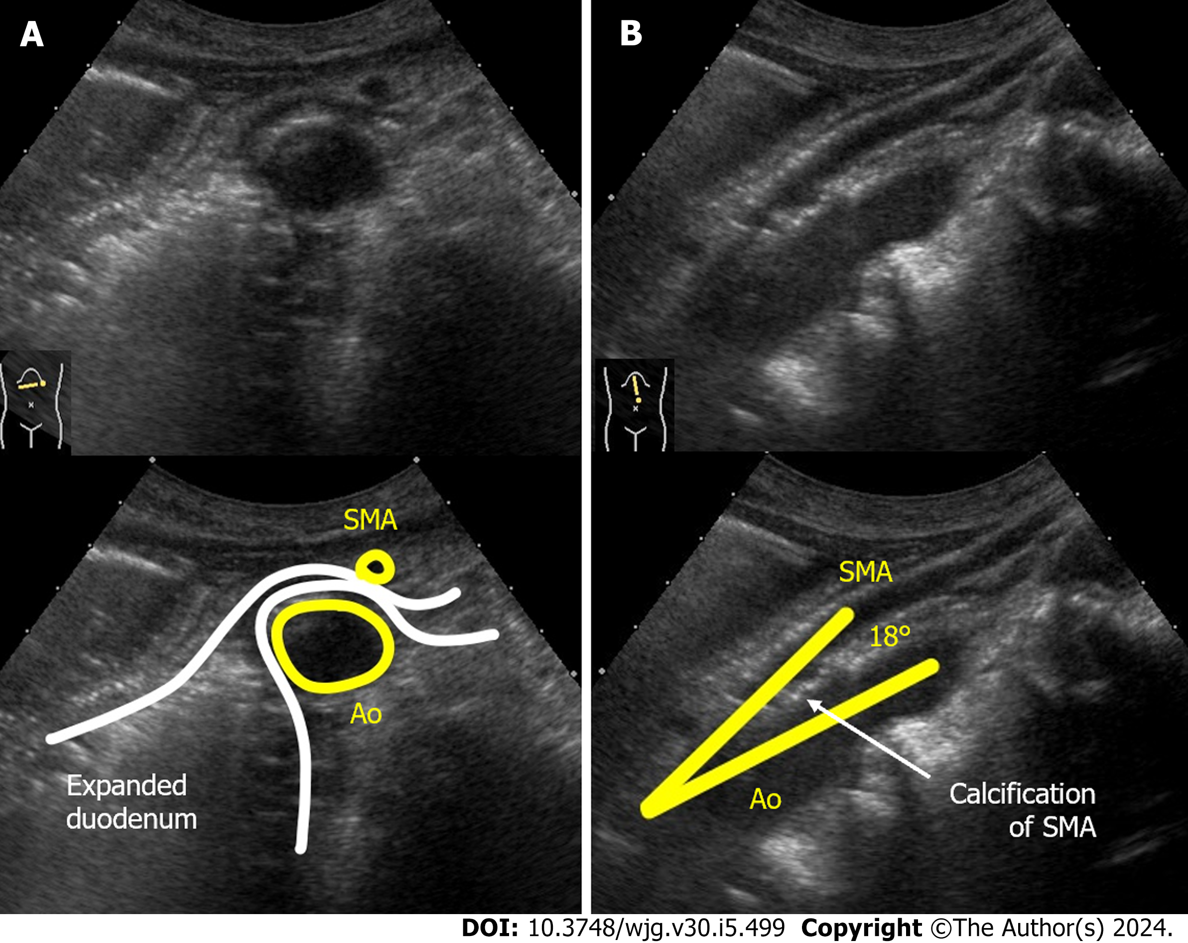
Figure 4 Dynamic ultrasonography images of case 2.
A: The supine position of ultrasonography confirmed expansion of the third part of the duodenal lumen and extrinsic compression of the duodenum by the superior mesenteric artery (SMA). The distance between the SMA and the aorta (Ao) was narrowed to 5 mm; B: Epigastric longitudinal scan showed that the angle of SMA-Ao decreased to 18°. Calcifications (arteriosclerosis) of the aorta and the root of SMA was also observed. SMA: Superior mesenteric artery; Ao: Aorta.
- Citation: Hasegawa N, Oka A, Awoniyi M, Yoshida Y, Tobita H, Ishimura N, Ishihara S. Dynamic ultrasonography for optimizing treatment position in superior mesenteric artery syndrome: Two case reports and review of literature. World J Gastroenterol 2024; 30(5): 499-508
- URL: https://www.wjgnet.com/1007-9327/full/v30/i5/499.htm
- DOI: https://dx.doi.org/10.3748/wjg.v30.i5.499


