Copyright
©The Author(s) 2024.
World J Gastroenterol. Feb 7, 2024; 30(5): 499-508
Published online Feb 7, 2024. doi: 10.3748/wjg.v30.i5.499
Published online Feb 7, 2024. doi: 10.3748/wjg.v30.i5.499
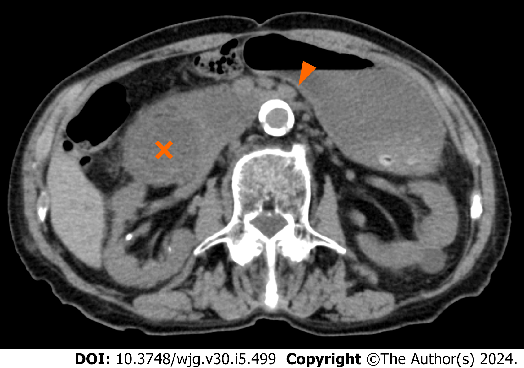
Figure 1 Computed tomography images of case 1.
The axial view of computed tomography showed the compression of the third portion of the duodenum between the superior mesenteric artery and the aorta (arrowhead) as well as markedly dilated duodenum (cross) and stomach.
- Citation: Hasegawa N, Oka A, Awoniyi M, Yoshida Y, Tobita H, Ishimura N, Ishihara S. Dynamic ultrasonography for optimizing treatment position in superior mesenteric artery syndrome: Two case reports and review of literature. World J Gastroenterol 2024; 30(5): 499-508
- URL: https://www.wjgnet.com/1007-9327/full/v30/i5/499.htm
- DOI: https://dx.doi.org/10.3748/wjg.v30.i5.499


