Published online May 7, 2017. doi: 10.3748/wjg.v23.i17.3084
Peer-review started: December 28, 2016
First decision: January 19, 2017
Revised: February 20, 2017
Accepted: March 21, 2017
Article in press: March 21, 2017
Published online: May 7, 2017
Processing time: 130 Days and 0.7 Hours
To report about the combination and advantages of a stapler-assisted diverticulotomy performed by flexible endoscopy.
From November 2014 till December 2015 17 patients (8 female, 9 male, average age 69.8 years) with a symptomatic Zenker diverticulum (mean size 3.5 cm) were treated by inserting a new 5 mm fully rotatable surgical stapler (MicroCutter30 Xchange, Cardica Inc.) next to an ultrathin flexible endoscope through an overtube. The Patients were under conscious sedation with the head reclined in left position, the stapler placed centrally and pushed forward to the bottom of the diverticulum. The septum was divided by the staple rows under flexible endoscopic control.
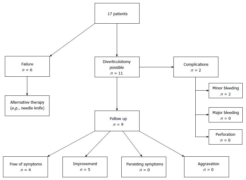
In eleven patients (64.7%) the stapler successfully divided the septum completely. Mean procedure time was 21 min, medium size of the septum was 2.8 cm (range 1.5 cm to 4 cm). In four patients the septum was shorter than 3 cm, in seven longer than 3 cm. To divide the septum, averagely 1.3 stapler cartridges were used. Two minor bleedings occurred. Major adverse events like perforation or secondary haemorrhage did not occur. After an average time of two days patients were discharged from the hospital. In 6 patients (35.3%) the stapler failed due to a thick septum or insufficient reclination of the head. Follow up endoscopy was performed after an average of two months in 9 patients; 4 patients (44.4%) were free of symptoms, 5 patients (55.6%) stated an improvement. A relapse of symptoms did not occur.
Flexible endoscopic Zenker diverticulotomy by using a surgical stapler is a new, safe and efficient treatment modality. A simultaneously tissue opening and occlusion prevents major complications.
Core tip: The new flexible MicroCutter30 XChange can dissect the septum of a Zenker diverticulum (ZD) under flexible endoscopic vision control. The simultaneous division and closure of the cutting edge by the staple rows prevents major bleedings or perforations. The combination of a flexible endoscopy and a surgical stapler is a significant advantage in the treatment of ZD.
- Citation: Wilmsen J, Baumbach R, Stüker D, Weingart V, Neser F, Gölder SK, Pfundstein C, Nötzel EC, Rösch T, Faiss S. New flexible endoscopic controlled stapler technique for the treatment of Zenker's diverticulum: A case series. World J Gastroenterol 2017; 23(17): 3084-3091
- URL: https://www.wjgnet.com/1007-9327/full/v23/i17/3084.htm
- DOI: https://dx.doi.org/10.3748/wjg.v23.i17.3084
Zenker’s diverticulum is an acquired pulsion diverticulum in an area of muscular weakness at the lateral posterior wall of the hypopharynx, the “Kilian triangle“. First described by Abraham Ludlow in 1764 it got formally named after the pathologist Friedrich Albert von Zenker in 1867[1]. It occurs more often in men than in women, usually between the seventh and eighth decade of life, the prevalence is ranging from 0.01% to 0.11%[2]. Although pathophysiology of its formation is not as yet completely understood, an increased luminal pressure is one of the main theories[3]. Because of the increased luminal pressure the mucosa and submucosa of the esophagus between the oblique fibers of the inferior pharyngeal constrictor and the horizontal fibers of the cricopharyngeus muscle pouches out[2]. Also besides the anatomical weakness of the Kilian triangle between the pharyngoesophageal muscles, a dysfunction of the upper esophageal sphincter (the cricopharyngeus muscle) with an increased muscle tone during swallowing is another hypothesis for the herniation[4].
With an increasing size of the diverticulum an initial globus can change into progressive dysphagia. Chronic cough, weight loss and aspiration of undigested food are other common symptoms. The surgical resection requires general anesthesia, complications like recurrent nerve palsy or haematoma occur more often and length of hospital stay (LOS) is significantly longer compared to endoscopic approaches[5], but 90 up to 95% of the patients are free of symptoms after open surgery[6].
Due to technical improvements less invasive endoscopic dissection of the cricopharyngeal bridge became more popular leading to a shorter LOS[7]. By dissecting the septum which includes muscular fibers of the cricopharyngeus and separates the diverticular sac from the esophageal lumen the pouch “collapses” and the two lumina become one. The septum can be dissected by either using a rigid endoscope (most common combined with a CO2 laser or a stapler) or a flexible endoscope[8]. Rigid endoscopy is performed under general anesthesia with the patient in supine position with overextended neck. To visualize the septum, usually a special rigid diverticuloscope, the spreadable Weerda diverticuloscope, is used. It consists of a short and a longer blade. The short blade intubates the diverticulum, the longer one the esophagus. Spreading the blades the septum gets distended allowing a better anatomic overview[9]. Stapling seems to be the preferred rigid approach based on a shorter LOS, a lower complication and lower failed exposure rate compared to laser diverticulotomy[5,10]. However, a significant disadvantage of the rigid endoscopy method is the requirement for general anesthesia, which might cause complications in elderly patients especially.
In 1995 Ishioka et al[11] published the first case series of a flexible endoscopic septum incision by using a needle knife. Compared to the surgical and rigid endoscopy treatment options, flexible endoscopy can be performed under conscious sedation, which is a major advantage. Up to now, different techniques and cutting devices are available to dissect the septum; free hand, guidewire/nasogastric tube, endoscopic cap or overtube assisted cut[12] with the use of argon plasma coagulation (APC), needle knives, endoscopic scissors or ESD knives[13-16]. It is still unclear which technique has the highest efficiency and safety and there is no standard treatment so far. The overall initial treatment success rate ranges from 56.4% to 100%, recurrence appears in a median of 10.5%[17]. Examining literature from 12 studies with 472 patients, the overall complication and mortality rate for flexible endoscopic Zenker’s diverticulotomy is 15% and 0% respectively. Most common complications are cervical emphysema (5.7%), perforation (4.0%) or bleeding (3.1%)[6].
The relapse of symptoms after flexible endoscopic treatment depends on whether the diverticular septum is divided completely or not. Trying to avoid a perforation the endoscopist might perform an incomplete dissection leading to a higher recurrence rate. On the other hand, dissecting too deep can increase the rate of cervical emphysema and the risk for mediastinitis. Therefore, using a stapler giving the possibility for a complete dissection of the septum combined with a simultaneously sealing of the wound edge seems to be an optimal solution especially when performed under flexible endoscopic control in conscious sedation as described first by Faiss et al[18].
The aim of this paper is to report about a case series demonstrating that the combination of a flexible endoscopy with a new 5 mm fully rotatable surgical stapler is a safe and efficient treatment modality in Zenker diverticulum (ZD).
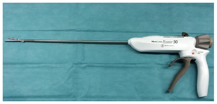
In seven different hospitals and one ambulant facility we performed a multicenter case series of a Zenker diverticulotomy by using a new 5 mm surgical stapler (MicroCutter30 Xchange, Cardica Inc., Redwood City, CA, United States/Cardica GmbH, Laichingen, Germany) (Figure 1). Besides the thin 5 mm shaft, the stapler articulates to 80 degrees in either direction. The cartridge consists of a 30 mm long staple line including 50 stainless steel staples. The stapler was developed for open or laparoscopic surgery procedures for example for the transection of the appendix.
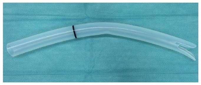
The patient was put under conscious sedation with the head reclined in left position. After performing a routine gastroscopy the stapler was introduced through a soft overtube (ZD overtube, Cook Endoscopy, Winston-Salem, NC, United States) (Figure 2), which was first placed over a standard endoscope and pushed forward up to approx. 20 cm from the teeth. Just like a spreadable diverticuloscope, the overtube consists of a short and a longer blade.
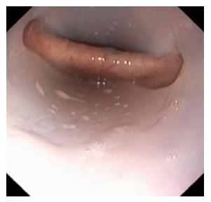
In correct position the short blade intubates the diverticulum, the longer the esophagus. The overtube stretches the septum and stabilizes the esophagus (Figure 3).
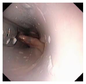
With the overtube in place, the standard endoscope was replaced by an ultrathin gastroscope (Olympus GIF XP 190 N; Olympus Optical Co., Europe, Hamburg, Germany). Because of the overtube’s 16 mm diameter and the 5 mm outer diameter of the endoscope, the 5 mm stapler can also be introduced through the overtube beside the endoscope. Under endoscopic control the stapler was opened, placed centrally and pushed forward to the bottom of the diverticulum with its shorter nose, whereas the longer nose (containing the stapler cartridge) was placed in the esophageal lumen (Figure 4).
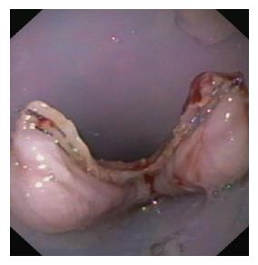
By pulling the trigger the stapler jaws clamp and simultaneously transect the septum between the two staple rows. If the septum of the ZD is endoscopically measured to be longer than 30 mm, the stapler can easily be unclamped, the used cartridge discarded, the stapler reloaded and the process is repeated towards the bottom of the diverticulum. After a complete dissection the stapler stitches are visible at the lateral rim of the divided septum of the ZD (Figure 5).
From November 2014 till October 2015 17 patients (8 female, 9 male) were treated by using this new flexible endoscopic controlled stapler technique. The only inclusion criterion was an already diagnosed, symptomatic ZD. A specified size of the diverticulum or a previous treatment were no exclusion criteria. After informing patients about possible complications (e.g., dental injuries, bleeding, perforation, mediastinitis, cardiovascular failure due to sedation), they gave their informed consent and data were collected by the participating endoscopists. The average age of the patients was 69.8 years (range 34-90 years), average female age was 72.9 years, average male age was 66.9 years (Table 1). Patients clinical symptoms were recorded before and 2 mo after the treatment.
| Total number of patients | n = 17 |
| Female | 8 (47.1) |
| Male | 9 (52.9) |
| Age, yr, mean (range) | 69.8 (34-90) |
| Mean female age, yr | 72.9 |
| Mean male age, yr | 66.9 |
| Mean size of ZD, cm (range) | 3.5 |
| Accumulated food in ZD | 5/17 (29.4) |
In 11 out of 17 patients (64.7%) septotomy was successfully performed. Mean procedure time was 21 min (range 10-45 min), medium size of the septum was 2.8 cm (range 1.5 cm to 4 cm). In four patients the septum was shorter than 3 cm, in seven longer than 3 cm. In five patients the ZD was stuffed with food. To divide the septum, averagely 1.3 stapler cartridges were used. In four patients two cartridges were required with a medium septum size of 3.25 cm.
Preventive application of antibiotics was not administered. Two minor bleedings occurred after septotomy and stopped after endoscopic placement of endoclips. No major adverse events like late onset bleeding or perforation occurred (Table 2). After the procedure, liquid intake was allowed, the day after oral feeding. Patients were discharged averagely two days after the procedure.
| Successful septotomy | Unsuccessful septotomy | |
| Number of cases | 11/17 (64.7%) | 6/17 (35.3%) |
| Procedure time, min, mean (range) | 21 (10-45) | 25 (10-50) |
| Septum size in cm, mean (range) | 2.8 (1.5-4) | 4.6 (3-10) |
| < 3 cm | n = 4 | n = 2 |
| > 3 cm | n = 7 | n = 4 |
| Used cartridges, mean | 1.3 | - |
In two patients (11.8%) the stapler could only partially divide the septum, due to steel staples placed in a previous rigid diverticulotomy and due to a very thick septum (stapler could only divide two-thirds of the tissue).
In four patients (23.5%) the stapler failed completely, caused by insufficient neck mobility (11.8%) and excessively thick tissue preventing the jaws from closing (11.8%) (Table 3). In these cases, the procedure was completed by needle knife dissection (Figure 6). Mean procedure time in uncomplete cases was 25 min (range 10-50 min), patients were also discharged averagely two days after the procedure.
| Failed stapler diverticulotomy | 6/17 (35.3) |
| Partial | 2/6 (11.8) |
| Complete | 4/6 (23.5) |
| Poor neck mobility | 2/4 (11.8) |
| Thick tissue | 2/4 (11.8) |
Follow up could be performed in 9 out of the 11 patients (81%) after two months in average. Two patients refused a follow up endoscopy. Four patients reported to be completely free of symptoms (dysphagia), five patients stated an improvement of symptoms.
Persistent symptoms or aggravation of symptoms could not be found. The septum was shortened from initial 2.8 cm to 0.6 cm in average. There was no more accumulation of food or liquid (Table 4). Up to now, no recurrence occurred.
| Total | 9/11 (81.8%) |
| Outcome | |
| Free of symptoms | n = 4 (44.4%) |
| Improved symptoms | n = 5 (55.6%) |
| Persisting symptoms | n = 0 |
| Aggravation | n = 0 |
| Septum size in cm, mean | |
| Before treatment | 2.8 |
| After treatment | 0.6 |
| Accumulated food in ZD | |
| Before treatment | 5/17 (29.4%) |
| After treatment | 0/9 |
Several different treatment modalities are available for symptomatic ZD. First, open surgery was increasingly replaced by rigid endoscopy. With the introduction of flexible endoscopes another treatment modality with a good clinical outcome, low invasivity and a short LOS became available. It is still under discussion which treatment should be used depending on age, diverticular size or comorbidities. Randomized trials are lacking, mostly the method is chosen by experience or personal preference. The tendency leads to flexible endoscopic procedures, open surgery remains an alternative in case of failed endoscopic exposure. A major advantage of flexible endoscopic treatment besides the low complication rate of 15% and mortality of 0% is that it can be performed under conscious sedation compared to surgery or rigid endoscopy[6]. Opponents of flexible endoscopy quote the high recurrence rate up to 35%[6], compared to a recurrence rate of 10% after a rigid stapler endoscopy[2] or 19% after surgical diverticular resection[19].
The higher recurrence rate after flexible endoscopic treatment could be caused by an incomplete dissection of the septum. Costamagna et al[20] demonstrated that the treatment success correlates with the length of the septotomy. The risk of perforation by cutting imprecisely and the difficulty to identify the last fibres of the cricopharyngeus could make the endoscopist keep a residual part of the septum to avoid cutting into the mediastinal space.
This is why Repici et al[16] used an ESD-knife to dissect the septum of 32 patients by hooking the tissue and pulling it up. The upward oriented tip helps to make the cut more precise, and thus the dissection of the last muscle fibres might be easier. With this new method no perforations occurred, but still two bleedings requiring APC for haemostasis. Also Brueckner et al[21] can report high success after using a hook-knife in 46 patients. All patients (100%) stated an initial clinical success after the treatment, recurrence appeared in 14 patients (30%) after a mean time of 4.4 mo. These patients were retreated (mean 1.39 sessions/patient) successfully except one patient. During the intervention in three patients arterial bleeding occurred and one patient developed a skin emphysema after the intervention.
The problem with all flexible cutting devices is that the dissected layers of the pouch do not get sealed during the division and therefore mediastinal emphysema could occur in 8% after flexible endoscopic treatment[5]. The prophylactical placement of endoclips at the bottom of the septum after the dissection is a described method to reduce this complication[22]. To avoid perforation, Li et al[23] performed a submucosal tunneling to cut the cricopharyngeal muscle fibers. The fibers were dissected by using a Hybrid knife, the mucosal incision was closed with endoclips. Symptoms resolved completely but the intervention required general anesthesia and the procedure was only performed in one patient so far.
In order to overcome these difficulties of conventional flexible endoscopic therapy we suggest that a stapling technique with a complete dissection of the septum together with a simultaneous closure of the cutting edge combined with the advantages of a flexible endoscopic approach seems to be an optimal treatment option in patients with symptomatic ZD. In our case series all successfully treated patients with the new endoscopic stapler method stated to be free of symptoms or showed convincing clinical improvement without any complication after two months.
However, there are some limitations of this new method: First: patients with an insufficient reclination of their neck could not be treated because the rigid stapler could not be pushed forward through the overtube to the septum. Second: In case of a very thick ZD septum the release of the stapler is blocked correctly for safety reasons. Therefore 6 out of 17 patients (35.3%) could not be treated by this innovative treatment modality. A preoperative imaging like X-ray, barium study or CT to provide information about the thickness of the diverticulum does not seem to be helpful. The evaluation of the thickness of the septum and if the stapler can be released is measured endoscopically during the diverticulotomy. In future these limitations will have to be overcome by a new curved stapler design with improved cramps enabling the dissection in patients also with insufficient neck reclination and/or thick septums in ZD.
In our case studies two minor bleedings occurred during the intervention which could be stopped endoscopically. We now believe that the bleedings would have stopped by themselves after removing the overtube. The overtube stretches the septum and generates tension, therefore haemostasis aggravates. Yuan et al[6] report a bleeding rate of 3.1% (examining 12 studies with 472 patients) after using a needle-knife/APC/monopolar forceps or a hook-knife for flexible endoscopic diverticulotomy.
Rieder et al[24] criticized that a rigid stapler diverticulotomy cannot be performed under endoscopic control due to the large 12 mm shaft of conventional staplers. Also generally after stapling a 10 mm long residual pouch remains because of the longer stapler tip. This problem does not appear after using our new MicroCutter30 Xchange; due to the slim 5 mm diameter of the shaft it is possible to place an ultrathin gastroscope simultaneously besides the stapler and dissect the septum under flexible endoscopic control. Also an only 5 mm long residual pouch remains because of the shorter 5 mm long tip of the used stapler.
Several authors claim small diverticula < 3 cm should not be treated endoscopically[25,26] and that the percentage of asymptomatic patients with small diverticula is higher after open procedures than after endoscopic treatment[27]. In small diverticula a complete dissection of the cricopharyngeus muscle up to 4 cm can be impossible. Because of a still remaining high intralumial pressure during swallowing a relapse can appear[7]. Also a correct septum exposure in small diverticula can be challenging caused by less instability and difficulty in placing the overtube. In contrast, in this small case series we cannot report a higher recurrence rate in patients with small diverticula or difficulty in placing the overtube. In the present case study the septum in four diverticula was shorter than 3 cm and there was no problem in the placement of the overtube or stapler. Neither can we confirm that the risk of perforation increases with large diverticula[28].
Another advantage of using a stapler for the septum dissection is a shorter procedure time (mean procedure time in our case study was 21 min) compared to other flexible techniques. Repici et al[29] for example required an average of 60 min for a flexible myotomy, Goelder et al[30] 32 min.
Concerning the costs of the used stapler device it is worth mentioning that the costs of this new stapler range between the costs of a conventional needle knife and a stag beetle knife (sb knife).
In conclusion our results show that the endoscopic treatment of ZD by using this new flexible controlled stapler technique is a simple and highly effective treatment modality with a very low complication rate. Further studies are needed with a modified, improved stapler enabling dissection of diverticula with a thicker septum tissue.
Flexible endoscopic septotomy is a well-established technique for the treatment of Zenker diverticulum. Various instruments including needle-knifes, hook-knifes, hot biopsy forceps, sb-knifes etc. are available to dissect the cricopharyngeal muscle. A major disadvantage of the various cutting devices is the creation of an unsealed and non-secured incision site, leading to increased complication rates such as major bleeding, oesophageal perforation and mediastinitis. Therefore we used a novel 5 mm surgical stapler in order to combine the advantages of a flexible endoscopy and a stapling technique.
Most recent published studies are reporting about new endoscopic knives/scissors for the dissection of the cricopharyngeal muscle. This is the first case study of a flexible stapler assisted Zenker diverticulotomy.
The combination of flexible endoscopy with the application of a surgical stapler has not yet been used before. The study showed that this new treatment modality is a safe, simple, fast and efficient method.
Using a stapler can lead to a shorter procedure time and a shorter length of hospital stay, based on the decreased bleeding risk and need for surveillance. The advantages of the combination may even enable the treatment in an ambulant setting.
The paper is written completely without any obvious problems. The number of patients is relatively low however just reports 1 year data.
Manuscript source: Invited manuscript
Specialty type: Gastroenterology and hepatology
Country of origin: Germany
Peer-review report classification
Grade A (Excellent): 0
Grade B (Very good): B
Grade C (Good): C, C
Grade D (Fair): 0
Grade E (Poor): 0
P- Reviewer: Cavalcoli F, Samiullah S, Zhang H S- Editor: Qi Y L- Editor: A E- Editor: Wang CH
| 1. | Haubrich WS. von Zenker of Zenker’s diverticulum. Gastroenterology. 2004;126:1269. [PubMed] |
| 2. | Law R, Katzka DA, Baron TH. Zenker’s Diverticulum. Clin Gastroenterol Hepatol. 2014;12:1773-1782; quiz 1773-1782. [RCA] [PubMed] [DOI] [Full Text] [Cited by in Crossref: 89] [Cited by in RCA: 91] [Article Influence: 8.3] [Reference Citation Analysis (0)] |
| 3. | Katzka DA, Baron TH. Transoral flexible endoscopic therapy of Zenker’s diverticulum: is it time for gastroenterologists to stick their necks out? Gastrointest Endosc. 2013;77:708-710. [RCA] [PubMed] [DOI] [Full Text] [Cited by in Crossref: 12] [Cited by in RCA: 13] [Article Influence: 1.1] [Reference Citation Analysis (0)] |
| 4. | Cook IJ, Gabb M, Panagopoulos V, Jamieson GG, Dodds WJ, Dent J, Shearman DJ. Pharyngeal (Zenker’s) diverticulum is a disorder of upper esophageal sphincter opening. Gastroenterology. 1992;103:1229-1235. [PubMed] |
| 5. | Verdonck J, Morton RP. Systematic review on treatment of Zenker’s diverticulum. Eur Arch Otorhinolaryngol. 2015;272:3095-3107. [RCA] [PubMed] [DOI] [Full Text] [Cited by in Crossref: 90] [Cited by in RCA: 92] [Article Influence: 8.4] [Reference Citation Analysis (0)] |
| 6. | Yuan Y, Zhao YF, Hu Y, Chen LQ. Surgical treatment of Zenker’s diverticulum. Dig Surg. 2013;30:207-218. [RCA] [PubMed] [DOI] [Full Text] [Cited by in Crossref: 108] [Cited by in RCA: 102] [Article Influence: 8.5] [Reference Citation Analysis (0)] |
| 7. | Ferreira LE, Simmons DT, Baron TH. Zenker’s diverticula: pathophysiology, clinical presentation, and flexible endoscopic management. Dis Esophagus. 2008;21:1-8. [RCA] [PubMed] [DOI] [Full Text] [Cited by in Crossref: 145] [Cited by in RCA: 136] [Article Influence: 8.0] [Reference Citation Analysis (0)] |
| 8. | Anagiotos A, Feyka M, Gostian AO, Lichtenstein T, Henning TD, Guntinas-Lichius O, Hüttenbrink KB, Preuss SF. Endoscopic laser-assisted diverticulotomy without versus with wound closure in the treatment of Zenker’s diverticulum. Eur Arch Otorhinolaryngol. 2014;271:765-770. [RCA] [PubMed] [DOI] [Full Text] [Cited by in Crossref: 2] [Cited by in RCA: 2] [Article Influence: 0.2] [Reference Citation Analysis (0)] |
| 9. | Papaspyrou G, Schick B, Papaspyrou S, Wiegand S, Al Kadah B. Laser surgery for Zenker’s diverticulum: European combined study. Eur Arch Otorhinolaryngol. 2016;273:183-188. [RCA] [PubMed] [DOI] [Full Text] [Cited by in Crossref: 8] [Cited by in RCA: 8] [Article Influence: 0.8] [Reference Citation Analysis (0)] |
| 10. | Miller FR, Bartley J, Otto RA. The endoscopic management of Zenker diverticulum: CO2 laser versus endoscopic stapling. Laryngoscope. 2006;116:1608-1611. [RCA] [PubMed] [DOI] [Full Text] [Cited by in Crossref: 49] [Cited by in RCA: 49] [Article Influence: 2.6] [Reference Citation Analysis (0)] |
| 11. | Ishioka S, Sakai P, Maluf Filho F, Melo JM. Endoscopic incision of Zenker’s diverticula. Endoscopy. 1995;27:433-437. [RCA] [PubMed] [DOI] [Full Text] [Cited by in Crossref: 123] [Cited by in RCA: 114] [Article Influence: 3.8] [Reference Citation Analysis (0)] |
| 12. | Costamagna G, Iacopini F, Tringali A, Marchese M, Spada C, Familiari P, Mutignani M, Bella A. Flexible endoscopic Zenker’s diverticulotomy: cap-assisted technique vs. diverticuloscope-assisted technique. Endoscopy. 2007;39:146-152. [RCA] [PubMed] [DOI] [Full Text] [Cited by in Crossref: 80] [Cited by in RCA: 76] [Article Influence: 4.2] [Reference Citation Analysis (0)] |
| 13. | Rabenstein T, May A, Michel J, Manner H, Pech O, Gossner L, Ell C. Argon plasma coagulation for flexible endoscopic Zenker’s diverticulotomy. Endoscopy. 2007;39:141-145. [RCA] [PubMed] [DOI] [Full Text] [Cited by in Crossref: 77] [Cited by in RCA: 85] [Article Influence: 4.7] [Reference Citation Analysis (0)] |
| 14. | Christiaens P, De Roock W, Van Olmen A, Moons V, D’Haens G. Treatment of Zenker’s diverticulum through a flexible endoscope with a transparent oblique-end hood attached to the tip and a monopolar forceps. Endoscopy. 2007;39:137-140. [RCA] [PubMed] [DOI] [Full Text] [Cited by in Crossref: 51] [Cited by in RCA: 58] [Article Influence: 3.2] [Reference Citation Analysis (0)] |
| 15. | Vogelsang A, Preiss C, Neuhaus H, Schumacher B. Endotherapy of Zenker’s diverticulum using the needle-knife technique: long-term follow-up. Endoscopy. 2007;39:131-136. [RCA] [PubMed] [DOI] [Full Text] [Cited by in Crossref: 67] [Cited by in RCA: 69] [Article Influence: 3.8] [Reference Citation Analysis (0)] |
| 16. | Repici A, Pagano N, Romeo F, Danese S, Arosio M, Rando G, Strangio G, Carlino A, Malesci A. Endoscopic flexible treatment of Zenker’s diverticulum: a modification of the needle-knife technique. Endoscopy. 2010;42:532-535. [RCA] [PubMed] [DOI] [Full Text] [Cited by in Crossref: 41] [Cited by in RCA: 40] [Article Influence: 2.7] [Reference Citation Analysis (0)] |
| 17. | Ishaq S, Hassan C, Antonello A, Tanner K, Bellisario C, Battaglia G, Anderloni A, Correale L, Sharma P, Baron TH. Flexible endoscopic treatment for Zenker’s diverticulum: a systematic review and meta-analysis. Gastrointest Endosc. 2016;83:1076-1089.e5. [RCA] [PubMed] [DOI] [Full Text] [Cited by in Crossref: 146] [Cited by in RCA: 119] [Article Influence: 13.2] [Reference Citation Analysis (0)] |
| 18. | Faiss S, Falck S, Cordruwisch W, Oldhafer KJ, Baumbach R. New flexible endoscopic controlled stapler technique for the treatment of Zenker’s diverticulum. Scand J Gastroenterol. 2015;50:1512-1515. [RCA] [PubMed] [DOI] [Full Text] [Cited by in Crossref: 3] [Cited by in RCA: 4] [Article Influence: 0.4] [Reference Citation Analysis (0)] |
| 19. | Antonello A, Ishaq S, Zanatta L, Cesarotto M, Costantini M, Battaglia G. The role of flexible endotherapy for the treatment of recurrent Zenker’s diverticula after surgery and endoscopic stapling. Surg Endosc. 2016;30:2351-2357. [RCA] [PubMed] [DOI] [Full Text] [Cited by in Crossref: 18] [Cited by in RCA: 25] [Article Influence: 2.5] [Reference Citation Analysis (0)] |
| 20. | Costamagna G, Iacopini F, Bizzotto A, Familiari P, Tringali A, Perri V, Bella A. Prognostic variables for the clinical success of flexible endoscopic septotomy of Zenker’s diverticulum. Gastrointest Endosc. 2016;83:765-773. |
| 21. | Brueckner J, Schneider A, Messmann H, Gölder SK. Long-term symptomatic control of Zenker diverticulum by flexible endoscopic mucomyotomy with the hook knife and predisposing factors for clinical recurrence. Scand J Gastroenterol. 2016;51:666-671. [RCA] [PubMed] [DOI] [Full Text] [Cited by in Crossref: 20] [Cited by in RCA: 24] [Article Influence: 2.7] [Reference Citation Analysis (0)] |
| 22. | Huberty V, El Bacha S, Blero D, Le Moine O, Hassid S, Devière J. Endoscopic treatment for Zenker’s diverticulum: long-term results (with video). Gastrointest Endosc. 2013;77:701-707. [RCA] [PubMed] [DOI] [Full Text] [Cited by in Crossref: 64] [Cited by in RCA: 70] [Article Influence: 5.8] [Reference Citation Analysis (0)] |
| 23. | Li QL, Chen WF, Zhang XC, Cai MY, Zhang YQ, Hu JW, He MJ, Yao LQ, Zhou PH, Xu MD. Submucosal Tunneling Endoscopic Septum Division: A Novel Technique for Treating Zenker’s Diverticulum. Gastroenterology. 2016;151:1071-1074. [RCA] [PubMed] [DOI] [Full Text] [Cited by in Crossref: 77] [Cited by in RCA: 100] [Article Influence: 11.1] [Reference Citation Analysis (0)] |
| 24. | Rieder E, Martinec DV, Dunst CM, Swanström LL. Flexible endoscopic Zenkers diverticulotomy with a novel bipolar forceps: a pilot study and comparison with needleknife dissection. Surg Endosc. 2011;25:3273-3278. [RCA] [PubMed] [DOI] [Full Text] [Cited by in Crossref: 18] [Cited by in RCA: 15] [Article Influence: 1.1] [Reference Citation Analysis (0)] |
| 25. | Jones D, Aloraini A, Gowing S, Cools-Lartigue J, Leimanis M, Tabah R, Ferri L. Evolving Management of Zenker’s Diverticulum in the Endoscopic Era: A North American Experience. World J Surg. 2016;40:1390-1396. [RCA] [PubMed] [DOI] [Full Text] [Cited by in Crossref: 16] [Cited by in RCA: 9] [Article Influence: 1.1] [Reference Citation Analysis (0)] |
| 26. | Rizzetto C, Zaninotto G, Costantini M, Bottin R, Finotti E, Zanatta L, Guirroli E, Ceolin M, Nicoletti L, Ruol A. Zenker’s diverticula: feasibility of a tailored approach based on diverticulum size. J Gastrointest Surg. 2008;12:2057-2064; discussion 2064-2065. [RCA] [PubMed] [DOI] [Full Text] [Cited by in Crossref: 58] [Cited by in RCA: 58] [Article Influence: 3.4] [Reference Citation Analysis (0)] |
| 27. | Gutschow CA, Hamoir M, Rombaux P, Otte JB, Goncette L, Collard JM. Management of pharyngoesophageal (Zenker’s) diverticulum: which technique? Ann Thorac Surg. 2002;74:1677-1682; discussion 1682-1683. [PubMed] |
| 28. | Buchanan MA, Riffat F, Mahrous AK, Fish BM, Jani P. Endoscopic or external approach revision surgery for pharyngeal pouch following primary endoscopic stapling: which is the favoured approach? Eur Arch Otorhinolaryngol. 2013;270:1707-1710. [RCA] [PubMed] [DOI] [Full Text] [Cited by in Crossref: 9] [Cited by in RCA: 9] [Article Influence: 0.7] [Reference Citation Analysis (0)] |
| 29. | Repici A, Pagano N, Fumagalli U, Peracchia A, Narne S, Malesci A, Rosati R. Transoral treatment of Zenker diverticulum: flexible endoscopy versus endoscopic stapling. A retrospective comparison of outcomes. Dis Esophagus. 2011;24:235-239. [RCA] [PubMed] [DOI] [Full Text] [Cited by in Crossref: 38] [Cited by in RCA: 37] [Article Influence: 2.6] [Reference Citation Analysis (0)] |
| 30. | Goelder SK, Brueckner J, Messmann H. Endoscopic treatment of Zenker’s diverticulum with the stag beetle knife (sb knife) - feasibility and follow-up. Scand J Gastroenterol. 2016;51:1155-1158. [RCA] [PubMed] [DOI] [Full Text] [Cited by in Crossref: 25] [Cited by in RCA: 22] [Article Influence: 2.4] [Reference Citation Analysis (0)] |


