Copyright
©The Author(s) 2016.
World J Gastroenterol. Sep 7, 2016; 22(33): 7518-7535
Published online Sep 7, 2016. doi: 10.3748/wjg.v22.i33.7518
Published online Sep 7, 2016. doi: 10.3748/wjg.v22.i33.7518
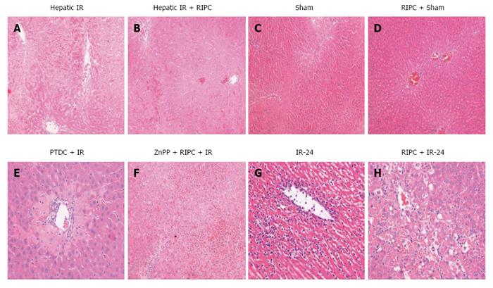
Figure 11 Hepatocytes showed vacuolation.
A: IR - the HE section shows large areas of necrosis and sinusoidal congestion, normal residual hepatocytes noted at bottom of the frame; B: RIPC + IR - The HE section shows sinusoidal congestion, some hepatocyte vacuolation but no significant necrosis; C: Sham - The HE section reveals no significant damage; D: RIPC + Sham - The HE section reveals congested central vein but no other significant change; E: PDTC + IR-diffuse congestion and patchy necrosis; F: ZNPP + RIPC + IR - extensive necrosis seen; G: Very severe injury with abundant ballooning degeneration and necrosis is seen in the IR-24 injury group. Very diffuse and significant neutrophil adhesion is seen in the IR group. Apoptosis is evident in the IR group; H: RIPC + IR-24 group shows less injury with some ballooning and degeneration as well as neutrophilic infiltration. RIPC: Remote ischemic preconditioning; IRI: Ischemia reperfusion injury; HO: Haemoxygenase.
- Citation: Tapuria N, Junnarkar S, Abu-amara M, Fuller B, Seifalian AM, Davidson BR. Haemoxygenase modulates cytokine induced neutrophil chemoattractant in hepatic ischemia reperfusion injury. World J Gastroenterol 2016; 22(33): 7518-7535
- URL: https://www.wjgnet.com/1007-9327/full/v22/i33/7518.htm
- DOI: https://dx.doi.org/10.3748/wjg.v22.i33.7518


