Published online Feb 21, 2015. doi: 10.3748/wjg.v21.i7.2220
Peer-review started: July 1, 2014
First decision: July 21, 2014
Revised: September 26, 2014
Accepted: November 30, 2014
Article in press: December 1, 2014
Published online: February 21, 2015
Processing time: 225 Days and 23.5 Hours
Multiple rectal carcinoids are rare. Due to the unreliability of endoscopic polypectomy in treating these submucosal lesions, a laparotomy is usually performed. We present a case report on multiple rectal carcinoids with three carcinoid foci < 10 mm in diameter located in the mid-rectum. Preoperative examination showed the lesions to be confined to the submucosal layer with no perirectal nodal involvement. A transanal endoscopic microsurgery was successfully performed to remove the three lesions with accurate full-thickness resection followed by secured suture closure. The postoperative pathology revealed neuroendocrine tumors G1 (carcinoids) located within the submucosal layer without lymphatic or vessel infiltration. Both the deep and lateral surgical margins were completely free of tumor cells. The patient recovered quickly and uneventfully. No tumor recurrence was observed at the six-month follow-up. For the multiple small rectal carcinoids without muscularis propria or lymphatic invasion, transanal endoscopic microsurgery offers a reliable and efficient alternative approach to traditional laparotomy for select patients, with the added advantages of minimally invasive surgery.
Core tip: A rare case of multiple small rectal carcinoids being successfully removed using the transanal endoscopic microsurgery technique. On the basis of careful preoperative evaluation and detailed postoperative pathological examination, transanal endoscopic microsurgery offers a safe, reliable and efficient alternative approach to the traditional surgeries for select patients with multiple rectal carcinoids.
- Citation: Zhou JL, Lin GL, Zhao DC, Zhong GX, Qiu HZ. Resection of multiple rectal carcinoids with transanal endoscopic microsurgery: Case report. World J Gastroenterol 2015; 21(7): 2220-2224
- URL: https://www.wjgnet.com/1007-9327/full/v21/i7/2220.htm
- DOI: https://dx.doi.org/10.3748/wjg.v21.i7.2220
With the rise in numbers of patients undergoing endoscopy screening, the detection rate of early rectal carcinoids has increased notably in recent years[1-4]. In most cases, rectal carcinoids occur in a singlet; however, multiple rectal carcinoids can occur and the reported incidence rates range from 2% to 4.5%[5-8]. The consensus is that small rectal carcinoids (≤ 10 mm), without muscularis propria invasion, can be treated curatively using local excision since they rarely metastasize[4,9,10]. Here, we present a case of multiple rectal carcinoids being successfully treated with local resection by transanal endoscopic microsurgery (TEM).
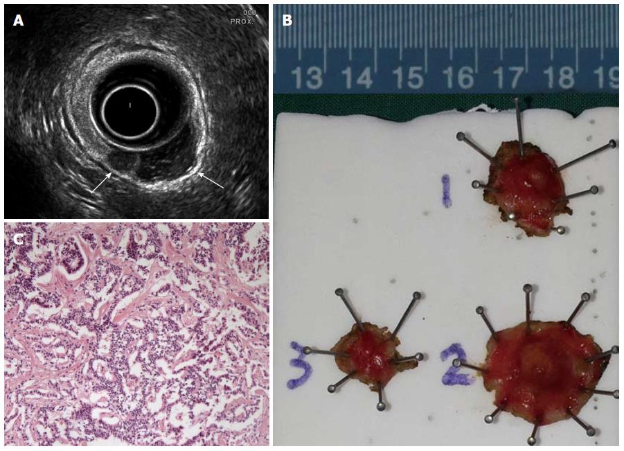
A 47-years-old Chinese male patient, presented with a year-long intermittent hematochezia, was admitted to our tertiary care center in October 2013. The signs of carcinoid syndrome were not observed. One month prior to his admission, the patient underwent a colonoscopy in a local hospital. The examination revealed three sessile nodules on the posterior wall of the rectum, 6 to 8 cm above the anal verge. The nodules, covered with yellowish discolored mucosa, 0.5 to 0.8 cm in diameter, were located within a relatively small area at 1 to 2 cm away from each other. During endoscopy, the smallest nodule was removed via snare polypectomy, and histological examination confirmed the diagnosis of rectal carcinoid. After one and a half months, the patient attended our clinic for further treatment. A transrectal ultrasound was performed which identified two hypoechoic nodules, 0.5 and 0.72 cm in maximum diameter, confined to the submucosal layer of the rectal wall (Figure 1A). No enlarged perirectal lymph node was observed. Hence, the initial diagnosis of multiple rectal carcinoids was made. Because of the small tumor size (< 10 mm) and the absence of muscularis propria invasion and nodal involvement, local excision was performed using the TEM technique.
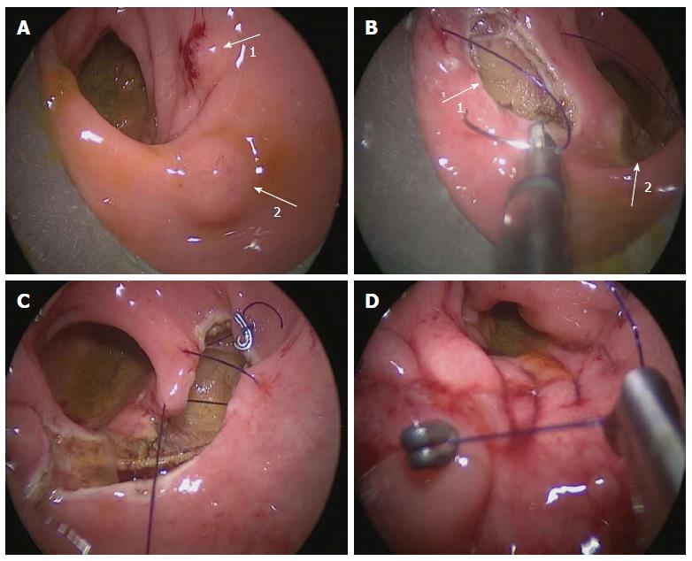
Rigid sigmoidoscopy was used to confirm locations of the three foci (including one scar site), followed by TEM under general anesthesia. The patient was placed in the lithotomy position with the lesions placed at the bottom of the operating field. The procedure was performed as previously described by Buess et al[11], using the Buess original TEM system (Richard Wolf GmbH, Knittlingen, Germany). After marking the resection area with coagulation dots by a needle cautery, ensuring a free margin area of 1 cm, the two submucosal tumors and the scar site of the third lesion were removed one at a time with full-thickness excision. Defects in the rectal wall were irrigated and closed using the running sutures of 3/0 absorbable monofilaments (Figure 2). The operation was completed within 40 min with a proximate blood loss of 10 mL.
No analgesic was required postoperatively. An elementary diet was initiated on the second day, and the patient was discharged two days after the surgery with an uneventful recovery. At the six-month follow-up, the patient was well with no carcinoid recurrence.
The postoperative pathology revealed two neuroendocrine tumors of 5 mm and 7 mm in diameter (specimen 1 and 2). In specimen 3, the residual neuroendocrine tumor cells were identified in the submucosal layer (Figure 1B and C). All three tumors were located within the submucosal layer with no lymphatic or vessel infiltration. Both the deep and lateral surgical margins were completely tumor-free in all specimens. Tumor cells showed a low cell proliferation index (Ki-67 < 2%). Immunohistochemistry study showed that tumor cells were positive for CD56 (NK-1) and Syn, and negative for CgA and p53 staining. The final diagnosis was multiple rectal neuroendocrine tumors G1 (carcinoids).
Multiple carcinoid tumors develop rarely in the rectum, with the incidence around 2% to 4.5%[5-8]. Sporadic reports of such cases can be retrieved from the literature written in the English language. Table 1 shows a summary of six multiple rectal carcinoids cases from five reports written in the English language. Among these six cases, four were extremely rare cases containing numerous endocrine cell micronests in the rectal wall[12-14], necessitating the radical resection with lymph node dissection. However, due to the small sample size (four out of six cases) it is not appropriate to conclude that multiple rectal carcinoids are often accompanied with numerous endocrine cell micronests.
Small rectal carcinoids (≤ 10 mm) without muscularis propria invasion can be curatively treated using local excision, due to the low metastasis rate[4,9,10]. Endoscopic polypectomy has been widely used for treatment of rectal carcinoids. However, most carcinoid tumors arise from the deep portion of the epithelial glands, penetrating the muscularis mucosa into the submucosal layer where the carcinoids form a nodular lesion[9]. Hence, the intrinsic limitations of the conventional endoscopic polypectomy result in a high chance of incomplete resection[9,15]. In the case of our patient, the postoperative pathologic result showed that lesion 3 had not been completely removed by endoscopic polypectomy. More advanced endoscopic resection methods, including endoscopic mucosal resection with cap, endoscopic submucosal resection with band ligation, and endoscopic submucosal dissection, or surgical local excision can be considered as more appropriate treatment methods[9].
Since its introduction by Buess et al[16] in 1983, TEM has emerged as an effective minimally invasive surgery for local resection of rectal lesions. With an average distance from the anal verge of about 7-8 cm[2,9], most small rectal carcinoids without metastasis are ideal candidates for local resection under TEM. This technique enables full-thickness excision and ensures accurate resection with sufficient margins by applying the delicate instruments under the superior visualization. In addition, TEM allows suturing of the rectal wall defects after tumor resection, thus securing sufficient excision without worrying about bowel perforation. In comparison with endoscopic resection methods, including advanced techniques of endoscopic mucosal resection with cap and endoscopic submucosal resection with band ligation, TEM enables a much larger extent of resection, ensuring more satisfactory oncological results for lesions with malignant potential.
In our case, three rectal carcinoids laid adjacent to each other within a relatively small area. It only took a small adjustment of the rectoscope to accomplish excision of all three lesions without changing the patient’s position. After removal of the tumors, adjacent defects were regarded as a whole and sutured together. The operation was, therefore, performed conveniently and efficiently using the TEM technique. However, for cases of multiple carcinoids containing numerous micronests of tumor cells in the rectum, an extremely rare condition[13,14], anterior resection or even abdominoperineal resection should be performed due to the inability of the naked eye to detect diffusely scattered tumor foci. For our patient, postoperative pathology excluded such a condition.
Although the majority of the small rectal carcinoids can be radically removed by local excision, some authors have reported that rectal carcinoids no larger than 10 mm may still spread to perirectal lymph nodes, with reported incidence rates of 7% to 9.7%[10,17]. Before making a local excision by TEM technique, we routinely perform the transrectal ultrasound to assess the depth of tumor invasion and evaluate the status of perirectal lymph nodes. Prior to surgery, a careful examination with rigid sigmoidoscopy is necessary to determine the exact location and orientation of the lesion, which is important for planning of the patient’s positioning during the surgery, allowing the lesion to sit at the 6-o’clock position and to facilitate the TEM operation.
Careful postoperative histological examination of specimens was required to evaluate the tumor size, the depth of invasion, the margin status, and the presence of risk factors of metastasis[2,10,18]. Tumors with invasion to muscularis propria, angiolymphatic invasion presence, or the increased indices of cell proliferation (such as Ki-67) should be treated with a salvage radical resection, or close follow-up should be performed.
To our knowledge, this is the first reported case of multiple rectal carcinoids being managed by TEM. In this case, high standard local tumor resection of multiple lesions was performed efficiently with minimal operative trauma; the patient recovered smoothly and quickly, further indicating the advantages of the TEM technique. Although this report presents only one case with a short follow-up period, we strongly believe that repeated TEM procedures for treating multiple rectal carcinoids will provide surgeons with invaluable experience, insight and knowledge of the disease and the best methods for treating this condition.
The patient was admitted with a year-long intermittent hematochezia.
The clinical diagnosis of multiple rectal carcinoids was made according to the result from colonoscopy that revealed three small submucosal nodules covered with yellowish-white mucosa on the rectal wall.
The small nodules on the rectal wall should be differentiated from the rectal adenomas, inflammatory polyps, gastrointestinal stromal tumors, lipomas, hamartomas, and rare diseases such as juvenile polyps, ganglioneuromatosis, etc.
Laboratory tests revealed no abnormal results.
The transrectal ultrasound showed small hypoechoic nodules being confined to the submucosal layer of the rectal wall.
Postoperative pathology revealed neuroendocrine tumors with a low cell proliferation index (Ki-67 < 2%).
The three lesions were surgically removed through local excisions using the transanal endoscopic microsurgery technique (TEM).
Five cases of multiple rectal carcinoids reported in articles written in the English language were reviewed and summarized.
For multiple small rectal carcinoids without muscularis propria or lymphatic invasion, TEM offers a reliable and efficient alternative approach to traditional laparotomy for select patients, with advantages of minimally invasive surgery. Careful preoperative evaluation and detailed postoperative pathological examination are mandatory to guarantee the radical removal of the tumor cells.
Most reviewers found this topic interesting. The manuscript was praised for being well-written and structured. Reviewers also recommended a summary of the previous reports on multiple rectal carcinoids.
P- Reviewer: Bernardin L, Fu W, Kasuga A, Messina F S- Editor: Gou SX L- Editor: A E- Editor: Liu XM
| 1. | Scherübl H. Rectal carcinoids are on the rise: early detection by screening endoscopy. Endoscopy. 2009;41:162-165. [RCA] [PubMed] [DOI] [Full Text] [Cited by in Crossref: 121] [Cited by in RCA: 135] [Article Influence: 8.4] [Reference Citation Analysis (0)] |
| 2. | Shields CJ, Tiret E, Winter DC. Carcinoid tumors of the rectum: a multi-institutional international collaboration. Ann Surg. 2010;252:750-755. [RCA] [PubMed] [DOI] [Full Text] [Cited by in Crossref: 112] [Cited by in RCA: 107] [Article Influence: 7.1] [Reference Citation Analysis (0)] |
| 3. | Taghavi S, Jayarajan SN, Powers BD, Davey A, Willis AI. Examining rectal carcinoids in the era of screening colonoscopy: a surveillance, epidemiology, and end results analysis. Dis Colon Rectum. 2013;56:952-959. [RCA] [PubMed] [DOI] [Full Text] [Cited by in Crossref: 48] [Cited by in RCA: 46] [Article Influence: 3.8] [Reference Citation Analysis (1)] |
| 4. | Kinoshita T, Kanehira E, Omura K, Tomori T, Yamada H. Transanal endoscopic microsurgery in the treatment of rectal carcinoid tumor. Surg Endosc. 2007;21:970-974. [RCA] [PubMed] [DOI] [Full Text] [Cited by in Crossref: 41] [Cited by in RCA: 35] [Article Influence: 1.9] [Reference Citation Analysis (0)] |
| 5. | BATES HR. Carcinoid tumors of the rectum. Dis Colon Rectum. 1962;5:270-280. [PubMed] |
| 6. | Caldarola VT, Jackman RJ, Moertel CG, Dockerty MB. Carcinoid tumors of the rectum. Am J Surg. 1964;107:844-849. [PubMed] |
| 7. | Quan SH, Bader G, Berg JW. Carcinoid tumors of the rectum. Dis Colon Rectum. 1964;7:197-206. [PubMed] |
| 9. | Son HJ, Sohn DK, Hong CW, Han KS, Kim BC, Park JW, Choi HS, Chang HJ, Oh JH. Factors associated with complete local excision of small rectal carcinoid tumor. Int J Colorectal Dis. 2013;28:57-61. [RCA] [PubMed] [DOI] [Full Text] [Cited by in Crossref: 54] [Cited by in RCA: 57] [Article Influence: 4.8] [Reference Citation Analysis (0)] |
| 10. | Konishi T, Watanabe T, Kishimoto J, Kotake K, Muto T, Nagawa H. Prognosis and risk factors of metastasis in colorectal carcinoids: results of a nationwide registry over 15 years. Gut. 2007;56:863-868. [RCA] [PubMed] [DOI] [Full Text] [Cited by in Crossref: 179] [Cited by in RCA: 179] [Article Influence: 9.9] [Reference Citation Analysis (0)] |
| 11. | Buess G, Kipfmüller K, Hack D, Grüssner R, Heintz A, Junginger T. Technique of transanal endoscopic microsurgery. Surg Endosc. 1988;2:71-75. [PubMed] |
| 12. | Kato M, Yonemura Y, Sugiyama K, Hashimoto T, Shima Y, Miyazaki I, Sugiura H, Kurumaya H, Hoso M, Yao T. [Multiple rectal carcinoids--with special reference to the histogenesis of these lesions]. Gan No Rinsho. 1986;32:1894-1900. [PubMed] |
| 13. | Maruyama M, Fukayama M, Koike M. A case of multiple carcinoid tumors of the rectum with extraglandular endocrine cell proliferation. Cancer. 1988;61:131-136. [PubMed] |
| 14. | Sasou S, Suto T, Satoh T, Tamura G, Kudara N. Multiple carcinoid tumors of the rectum: report of two cases suggesting the origin of carcinoid tumors. Pathol Int. 2012;62:699-703. [RCA] [PubMed] [DOI] [Full Text] [Cited by in Crossref: 10] [Cited by in RCA: 11] [Article Influence: 1.0] [Reference Citation Analysis (0)] |
| 15. | Jeon SM, Cheon JH. Rectal carcinoid tumors: pitfalls of conventional polypectomy. Clin Endosc. 2012;45:2-3. [RCA] [PubMed] [DOI] [Full Text] [Full Text (PDF)] [Cited by in Crossref: 4] [Cited by in RCA: 3] [Article Influence: 0.2] [Reference Citation Analysis (0)] |
| 16. | Buess G, Theiss R, Hutterer F, Pichlmaier H, Pelz C, Holfeld T, Said S, Isselhard W. [Transanal endoscopic surgery of the rectum - testing a new method in animal experiments]. Leber Magen Darm. 1983;13:73-77. [PubMed] |
| 17. | Soga J. Early-stage carcinoids of the gastrointestinal tract: an analysis of 1914 reported cases. Cancer. 2005;103:1587-1595. [RCA] [PubMed] [DOI] [Full Text] [Cited by in Crossref: 234] [Cited by in RCA: 220] [Article Influence: 11.0] [Reference Citation Analysis (0)] |
| 18. | Kasuga A, Chino A, Uragami N, Kishihara T, Igarashi M, Fujita R, Yamamoto N, Ueno M, Oya M, Muto T. Treatment strategy for rectal carcinoids: a clinicopathological analysis of 229 cases at a single cancer institution. J Gastroenterol Hepatol. 2012;27:1801-1807. [RCA] [PubMed] [DOI] [Full Text] [Cited by in Crossref: 56] [Cited by in RCA: 60] [Article Influence: 4.6] [Reference Citation Analysis (0)] |
| 19. | Okamoto Y, Fujii M, Tateiwa S, Sakai T, Ochi F, Sugano M, Oshiro K, Masai K, Okabayashi Y. Treatment of multiple rectal carcinoids by endoscopic mucosal resection using a device for esophageal variceal ligation. Endoscopy. 2004;36:469-470. [RCA] [PubMed] [DOI] [Full Text] [Cited by in Crossref: 25] [Cited by in RCA: 22] [Article Influence: 1.0] [Reference Citation Analysis (0)] |
| 20. | Haraguchi M, Kinoshita H, Koori M, Tsuneoka N, Kosaka T, Ito Y, Furui J, Kanematsu T. Multiple rectal carcinoids with diffuse ganglioneuromatosis. World J Surg Oncol. 2007;5:19. [RCA] [PubMed] [DOI] [Full Text] [Full Text (PDF)] [Cited by in Crossref: 21] [Cited by in RCA: 23] [Article Influence: 1.3] [Reference Citation Analysis (0)] |


