Copyright
©The Author(s) 2015.
World J Gastroenterol. Jan 14, 2015; 21(2): 644-652
Published online Jan 14, 2015. doi: 10.3748/wjg.v21.i2.644
Published online Jan 14, 2015. doi: 10.3748/wjg.v21.i2.644
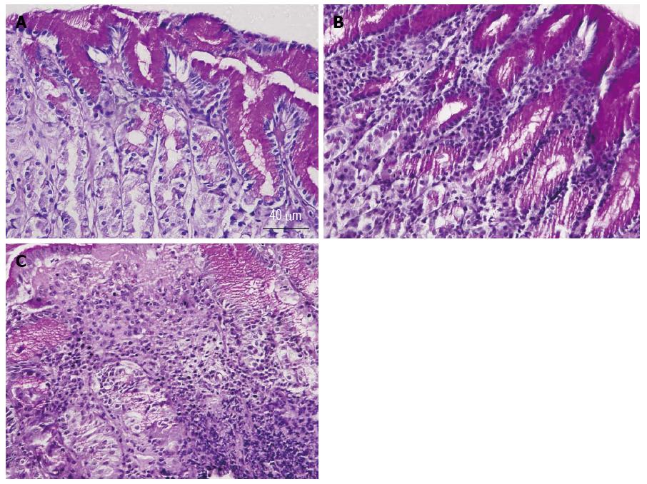
Figure 1 Representative micrographs of gastric corpus mucosal sections showing normal mucosa (A), superficial gastritis (B), and chronic atrophic gastritis (C).
Note the mild infiltration of the gastric mucosa by lymphoid cells near the luminal surface in superficial gastritis (B) and the massive infiltration of the mucosa by lymphoid cells in atrophic gastritis (C).
- Citation: Alfazari AS, Al-Dabbagh B, Al-Dhaheri W, Taha MS, Chebli AA, Fontagnier EM, Koutoubi Z, Kochiyi J, Karam SM, Souid AK. Profiling cellular bioenergetics, glutathione levels, and caspase activities in stomach biopsies of patients with upper gastrointestinal symptoms. World J Gastroenterol 2015; 21(2): 644-652
- URL: https://www.wjgnet.com/1007-9327/full/v21/i2/644.htm
- DOI: https://dx.doi.org/10.3748/wjg.v21.i2.644


