Published online Dec 14, 2014. doi: 10.3748/wjg.v20.i46.17670
Revised: June 16, 2014
Accepted: July 16, 2014
Published online: December 14, 2014
Processing time: 233 Days and 7.5 Hours
Splenic rupture is a common consequence of blunt abdominal trauma. Emergency splenectomy is indicated when conservative management is not effective. With better understanding of the immunologic function of the spleen, surgeons have begun to perform the splenic-preserving surgery. However, it is technical challenge to perform emergency laparoscopic partial splenectomy for patient with spleen rupture. A 15-year-old male patient suffered from grade III spleen injury basing on the American association for the surgery of trauma splenic injury scale. Conservative treatment failed to success basing on the dramatically decreased hemoglobin level. During the laparoscopic exploration, we found that two individual ruptures were associated with the upper pole of spleen. An emergency laparoscopic partial splenectomy was successfully carried out. The operative time was approximate 150 min and the estimated blood loss was 200 mL. The post-operative course was uneventful and the patient was discharged on the 7th post-operative day.
Core tip: It is technical challenge to perform laparoscopic partial splenectomy for patient with spleen rupture because of fragile parenchyma and abundant blood supply. We report a case of successfully laparoscopic partial splenectomy for ruptured spleen, with ordinary laparoscopic instruments. We also shared our experience in laparoscopic partial splenectomy in this case report.
- Citation: Cai YQ, Li CL, Zhang H, Wang X, Peng B. Emergency laparoscopic partial splenectomy for ruptured spleen: A case report. World J Gastroenterol 2014; 20(46): 17670-17673
- URL: https://www.wjgnet.com/1007-9327/full/v20/i46/17670.htm
- DOI: https://dx.doi.org/10.3748/wjg.v20.i46.17670
Splenic rupture is a common consequence of blunt abdominal trauma, which requiring emergency splenectomy when conservative management is not effective[1]. Generally, emergency open total splenectomy is required in this situation. Since Delaitre et al[2] firstly reported laparoscopic splenectomy (LS), LS has become the gold standard procedure for remove of normal to moderate enlarged spleens[3]. However, due to difficulty in blood control and poor visibility, it difficult to perform splenectomy for ruptured spleens laparoscopically[4].
With better understanding of immunologic function of the spleen, surgeons began to perform the splenic-preserving surgery during the last decades. Laparoscopic partial splenectomy (LPS) was performed for patients with benign splenic tumors by several skilled laparoscopic surgeons[5,6]. To our knowledge, LPS for ruptured spleen was rarely reported in the literature. Herein, we reported a case of emergency LPS for a patient who suffered from splenic rupture.
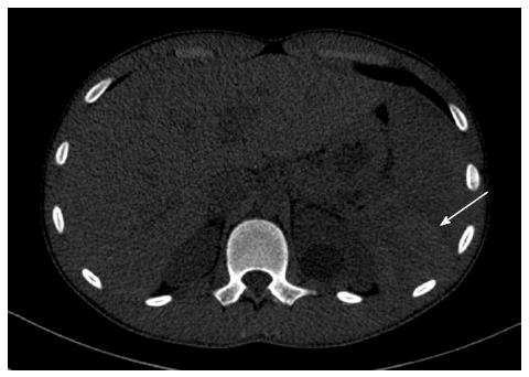
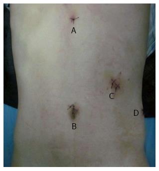
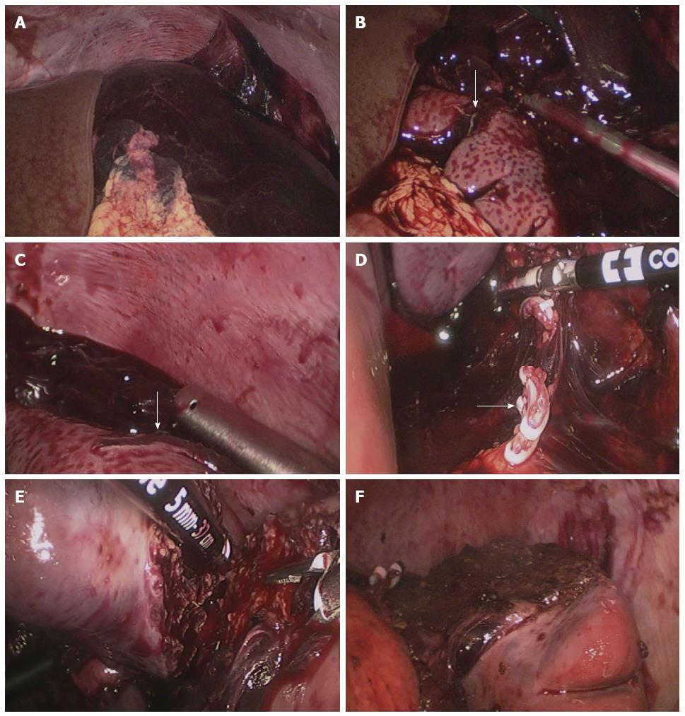
A 15-year-old male patient admitted at our institution following blunt abdominal trauma during skating. The computed tomography showed large hemoperitoneum associated with a traumatic rupture at the upper pole of the spleen (Figure 1). The patient suffered from grade III spleen injury basing on the American association for the surgery of trauma splenic injury scale. The hemoglobin decreased from 122 to 100 g/L during the hospitalization with four hours conservative treatment. The heart rate of the patient increased from 105 beats/min to 132 beats/min, and the blood pressure decreased from 130/83 mmHg to 97/56 mmHg. The grade of American Society of Anesthesiologists scale of this patient was grade 3. A laparoscopic partial splenectomy/splenectomy was scheduled with the consent of the patient’s parents. The patient received general anesthesia and was placed in the right semidecubitus position with the left side elevated approximately 60° and the operating table slightly tilted to the reverse Trendelenburg position. The sizes and distributions of trocars were shown in Figure 2. During laparoscopic exploration, a large hemoperitoneum (Figure 3A) and two individual ruptures of spleen were found (Figure 3B and C). Active bleeding was also indentified after removing the blood clot covering the ruptures. The intra-abdominal blood was salvage for autotransfusion after assurance of no liver rupture or hollow organs rupture. The operation began with clearance of hemoperitoneum, followed by dissection of splenogastric ligament (including the short gastric artery). In order to control the bleeding, we quickly controlled the splenic artery branches supplying the upper pole of spleen (Figure 3D). Then we dissected the splenic artery branches one by one until the ischemia line exceeding the rupture of the spleen. We dissected the upper pole of spleen parenchyma using a Ligasure system (Figure 3E). The oozing of blood from the cut edge was stopped by bipolar coagulator (Figure 3F). The dissected spleen tissue was putted into a retrieval bag, morcellated with forceps and retrieved from the 12-mm incision. A closed drainage was placed in the splenic fossa. The operative time was 150 min and the estimated blood loss was 200 mL. The post-operative course was uneventful.
Conservative managements are considered the current standard of care for hemodynamically stable patients with splenic injuries[7,8]. Emergency splenectomy is mandatory only when the patient suffers from low systolic blood pressure which does not increase after fluid resuscitation and blood transfusions[8]. The computed tomography (CT) scan has proven to be an excellent imaging modality for accurate diagnosis of splenic injury and its severity[7]. Repeated hemoglobin examination and ultrasonography are useful strategies to estimate the blood loss and detect the active bleeding. In this case, CT revealed a rupture and a large hematoma located at the posterior of spleen, where impeded us to perform splenorrhaphy. The hemoglobin level of this patient was 122 g/L on arrival. After four hours conservative therapy, the hemoglobin level decreased to 100 g/L. Laparoscopic partial splenectomy was required considering the active bleeding and the location of rupture.
It is a common consensus that laparoscopic surgery has several advantages over open surgery, such as better cosmetics, less blood loss, and faster recovery. However, due to difficulty in bleeding control and poor visibility associated with laparoscopic splenectomy, open surgery is the first choice in most of institutions in setting of splenic rupture. Owing to advancement in the operative instruments and accumulating of operative experience in laparoscopic splenectomy, several surgeons began to perform laparoscopic splenectomy for patients with ruptured spleens[1,4,8]. Huscher et al[8] report the largest series of patients treated laparoscopically for a splenic injury, including six cases of splenectomy, one case of upper polar resection, three cases of polyglycolic mesh wrapping, and one case of splenic hemostasis using argon beam coagulation. They concluded that it is technical feasible to perform laparoscopic splenic surgery for ruptured spleen. With better understanding of immunologic function of the spleen and the potential threat of severe post-splenectomy infections, surgeons began to pay more attention to parenchyma-preserving surgical procedures. In setting of splenic rupture, partial splenic artery embolization, radiofrequency ablation, splenorrhaphy, and partial splenectomy are available strategies to conserve the splenic parenchyma. Proper procedure should be chose basing on the location and severity of the rupture and the experience of the surgeon.
Because of fragile parenchyma and abundant blood supply, it is much more difficult to perform LPS than laparoscopic splenectomy. Poulin et al[9] reported the first case of LPS for ruptured spleen in 1995. They performed partial splenic artery embolism to control bleeding before the operation. Since then, this procedure has been very rarely reported due to technical challenge. Dr Peng, an experienced laparoscopic surgeon in China, has performed approximately 400 cases of laparoscopic splenectomy and 13 cases of LPS. Basing on our experience, several aspects are very important to carry out LPS for patients with ruptured spleen. The basic requirement for LS for ruptured spleen is that the patient should be hemodynamically stable. The CT scan shows the rupture locates at the upper pole or lower pole of spleen. During the operation, we should not remove blood clot covering the rupture as it is helpful to stop the bleeding. We dissected the branches of splenic artery until the ischemic demarcation line crossed the rupture and dissected splenic parenchyma 6 to 10 mm above the ischemic demarcation line. The oozing of blood from cut edge can be stopped by bipolar coagulator. The autotransfusion is also very important to patient with great blood loss; however, we should avoid its application in patients with hollow organ rupture or hepatic rupture. A closed drainage is required to monitor the probable bleeding from the stump of spleen. Basing on our limited experience, LPS is indicated for hemodynamically stable patients who fail to response to conservative treatment. The rupture of spleen is severe and not feasible to perform splenorrhaphy or other strategies. The location of rupture should be the upper pole or lower pole of spleen and the experience of surgeon is also critical. Due to unavailable in the literature, it should be stated that these recommendations were based on our own experience other than literature review or an objective study. Further studies are required to establish the selection criteria for LPS in setting of splenic rupture.
In conclusion, laparoscopic partial splenectomy for hemodynamically stable patient with splenic rupture is feasible. However, it is just suitable for selected patients and requires adequate experience with elective laparoscopic partial splenectomy.
Thanks to Dr. Ali Mohanmad Wajid for language editing.
A 15-year-old male patient suffered from grade III spleen injury basing on the American association for the surgery of trauma splenic injury scale.
The diagnosis of splenic rupture can be established basing on the history of injury and computed tomography (CT) image.
The repeated blood count examination revealed that hemoglobin decreased from 122 to 100 g/L during the hospitalization with four hours conservative treatment.
The CT image revealed that the patient suffered from grade III spleen injury located at the upper pole of spleen.
Emergency laparoscopic partial splenectomy.
To date, there was only one case of laparoscopic partial splenectomy in the literature.
Laparoscopic partial splenectomy for hemodynamically stable patients with a ruptured spleen is feasible and can preserve the immune function of spleen.
This is an interesting case report on ruptured spleen successfully treated by laparoscopic partial splenectomy. The authors provide selection criteria for the choice of laparoscopic partial splenectomy in the emergency situation basing on their own experience.
P- Reviewer: Chiu CC, Giannopoulos GA, Ikuta S, Maleki AR, Shimi SM S- Editor: Gou SX L- Editor: A E- Editor: Ma S
| 1. | Basso N, Silecchia G, Raparelli L, Pizzuto G, Picconi T. Laparoscopic splenectomy for ruptured spleen: lessons learned from a case. J Laparoendosc Adv Surg Tech A. 2003;13:109-112. [RCA] [PubMed] [DOI] [Full Text] [Cited by in Crossref: 23] [Cited by in RCA: 31] [Article Influence: 1.4] [Reference Citation Analysis (0)] |
| 2. | Delaitre B, Maignien B. [Splenectomy by the laparoscopic approach. Report of a case]. Presse Med. 1991;20:2263. [PubMed] |
| 3. | Silecchia G, Boru CE, Fantini A, Raparelli L, Greco F, Rizzello M, Pecchia A, Fabiano P, Basso N. Laparoscopic splenectomy in the management of benign and malignant hematologic diseases. JSLS. 2006;10:199-205. [PubMed] |
| 4. | Ren CJ, Salky B, Reiner M. Hand-assisted laparoscopic splenectomy for ruptured spleen. Surg Endosc. 2001;15:324. [RCA] [PubMed] [DOI] [Full Text] [Cited by in Crossref: 17] [Cited by in RCA: 18] [Article Influence: 0.8] [Reference Citation Analysis (0)] |
| 5. | Corcione F, Cuccurullo D, Caiazzo P, Settembre A, Bruzzese G, Vittoria I, Cusano T. Laparoscopic partial splenectomy for a splenic pseudocyst. Surg Endosc. 2003;17:1850. [RCA] [PubMed] [DOI] [Full Text] [Cited by in Crossref: 15] [Cited by in RCA: 12] [Article Influence: 0.6] [Reference Citation Analysis (0)] |
| 6. | Szczepanik AB, Meissner AJ. Partial splenectomy in the management of nonparasitic splenic cysts. World J Surg. 2009;33:852-856. [RCA] [PubMed] [DOI] [Full Text] [Cited by in Crossref: 32] [Cited by in RCA: 37] [Article Influence: 2.3] [Reference Citation Analysis (0)] |
| 7. | Uranüs S, Pfeifer J. Nonoperative treatment of blunt splenic injury. World J Surg. 2001;25:1405-1407. [PubMed] |
| 8. | Huscher CG, Mingoli A, Sgarzini G, Brachini G, Ponzano C, Di Paola M, Modini C. Laparoscopic treatment of blunt splenic injuries: initial experience with 11 patients. Surg Endosc. 2006;20:1423-1426. [RCA] [PubMed] [DOI] [Full Text] [Cited by in Crossref: 28] [Cited by in RCA: 30] [Article Influence: 1.6] [Reference Citation Analysis (0)] |
| 9. | Poulin EC, Thibault C, DesCôteaux JG, Côté G. Partial laparoscopic splenectomy for trauma: technique and case report. Surg Laparosc Endosc. 1995;5:306-310. [PubMed] |


