Published online Sep 15, 1996. doi: 10.3748/wjg.v2.i3.144
Revised: July 4, 1996
Accepted: August 10, 1996
Published online: September 15, 1996
AIM: To analyze the radiological features of ulcerative diseases of the small bowel.
METHODS: Thirty-five patients (20 men and 15 women) with inflammatory ulcerative bowel diseases were studied by radiography (barium meal and/or double contrast study). Patient diseases included eleven cases of tuberculosis (TB), thirteen cases of Crohn’s disease, seven cases of bowel Behcet disease, two cases of simple ulcers, and two cases of ischemic bowel disease. Diagnosis was established pathologically in 33 cases and by clinical observation after therapy in two cases.
RESULTS: The lesions were located in the ileum of 82% of TB cases, 77% of Crohn’s disease cases, 71% of bowel Behcet disease cases, 50% of simple ulcer cases, and 100% of ischemic bowel disease cases. Ulceration was always present with variable appearances. Longitudinal ulcers, and fissures were noted in Crohn’s disease only. There were five cases of large and deep ulcers, three of which were bowel Behcet disease. Superficial and irregular ulcers were present in ten TB cases, and , and transverse ulcers were identified in two TB cases.
CONCLUSION: The morphological appearances of the ulcer, surrounding mucosal alterations, and bowel deformation were the basis for the radiological diagnosis. Correct diagnosis was dependent on optimal X-ray examination techniques and proper interpretation of the morphological changes.
- Citation: Lu Y, Duan JY, Gao Y. Radiological diagnosis of inflammatory ulcerative diseases of the small bowel. World J Gastroenterol 1996; 2(3): 144-145
- URL: https://www.wjgnet.com/1007-9327/full/v2/i3/144.htm
- DOI: https://dx.doi.org/10.3748/wjg.v2.i3.144
Intestinal ulcers are often found in cases of small intestine inflammatory disease. They are accompanied by a high mortality rate.
It is difficult to make differential diagnoses by radiographic examination[1,2]. In this study, we reviewed 35 cases of small intestine disease, and analyzed the radiographic characteristics of the disease. Serial changes in radiographic examination were observed.
We performed radiography (barium meal and/or double contrast study) in 35 patients (20 men and 15 women). All patients were diagnosed with small intestine inflammation with an intestinal ulcer. Diagnosis was established by pathology in 33 cases and by clinical observation after therapy in two cases. The age range of the patients was 14 years to 78 years. Most patients had gastrointestinal symptoms such as abdominal pain, diarrhea, and bloody stool. The inflammatory ulcerative bowel diseases included eleven cases of tuberculosis (TB), thirteen cases of Crohn’s disease, seven cases of bowel Behcet disease, two cases of simple ulcers, and two cases of ischemic bowel disease.
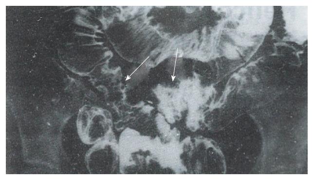
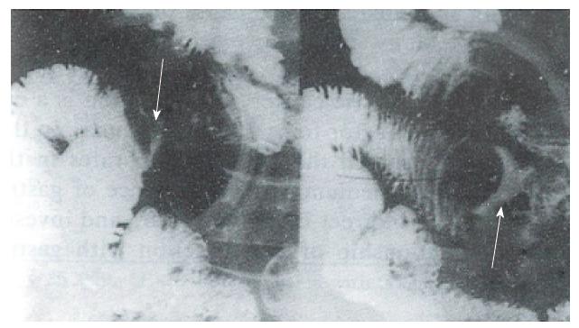
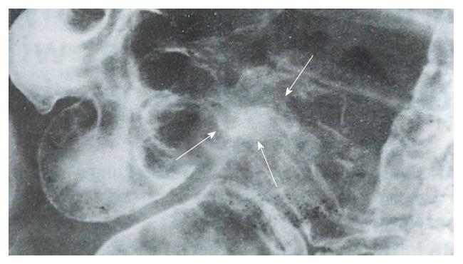
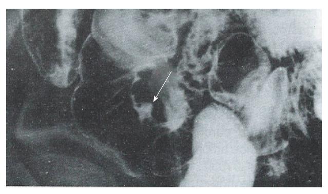
The lesions were located in the ileum of 82% of TB cases, of 77% of Crohn’s disease cases, of 71% of bowel Behcet disease cases, of 50% of simple ulcer cases, and of 100% of ischemic bowel disease cases. Ulceration was always present with variable appearances. Longitudinal ulcers and fissures were noted in Crohn’s disease only. There were 5 cases of large and deep ulcers, three of which occurred in bowel Behcet disease. Superficial and irregular ulcers were present in ten TB cases, and transverse ulcers were observed in two TB cases (Figure 1 and Figure 2).
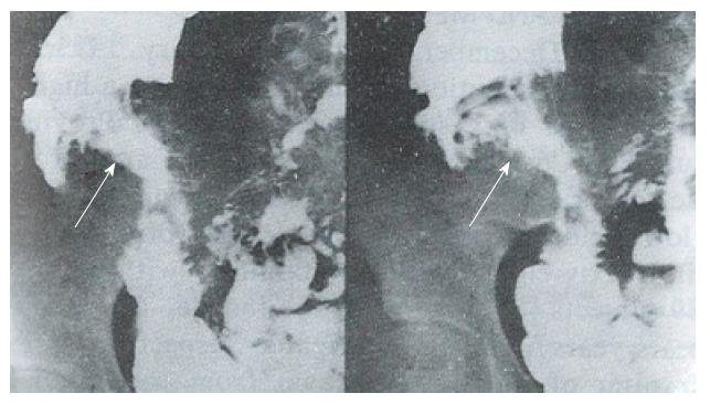
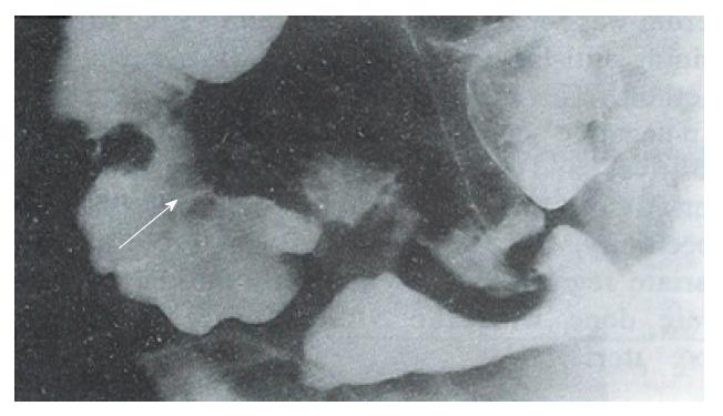
Ulceration is present in the majority of small bowel inflammatory diseases including TB, Crohn’s disease, bowel Behcet disease, simple ulcers, and ischemic bowel disease. The morphological appearances of the ulcer, surrounding mucosal alterations and bowel deformation were the basis for radiological diagnosis[3-9]. TB was located in the terminal ileum. Ulcerations had variable appearances, but no longitudinal ulcers and fissures were noted. In Crohn’s disease, the above characteristics were noted. In addition, cobblestone and fistulas occurred frequently (Figure 3 and Figure 4). The radiographic findings of bowel Behcet disease and simple ulcer were similar. However, the ulcers in bowel Behcet disease tended to be larger and deeper with surrounding mucosal edema (Figure 5 and Figure 6). All patients had mucocutaneous ocular symptoms. Ulcers in ischemic bowel disease had no characteristics (Figure 7). Correct diagnosis was dependent on optimal X-ray examination techniques and proper interpretation of the morphological changes. Enteroclysis, a controlled infusion method offering a double contrast and highly detailed examination of the small bowel, is relatively easy to perform. Despite the INTRODUCTION of enteroclysis, the peroral small bowel examination remains the predominant radiographic method of imaging the small intestine. This report emphasizes the technical factors needed to produce excellent peroral small bowel examinations.
Original title:
S- Editor: Ma JY L- Editor: Filipodia E- Editor: Li RF
| 1. | Feczko PJ, Holpert RD. Radiology of inflammatory bowel disease. Radiol Clin North Am. 1987;25:1-20. |
| 2. | Xie JX. Diagnosis and Differential Diagnosis of inflammatory ulcerative diseases of the small bowel. Zhonghua Fangshexue Zazhi. 1994;28:128-130. |
| 3. | Vaidya MG, Sodhi JS. Gastrointestinal tract tuberculosis: a study of 102 cases including 55 hemicolectomies. Clin Radiol. 1978;29:189-195. [RCA] [PubMed] [DOI] [Full Text] [Cited by in Crossref: 40] [Cited by in RCA: 39] [Article Influence: 0.8] [Reference Citation Analysis (0)] |
| 4. | Yao T, Okada M, Fuchigami T, Iida M, Takenaka K, Date H, Fujita K. The relationship between the radiological and clinical features in patients with Crohn’s disease. Clin Radiol. 1989;40:389-392. [RCA] [PubMed] [DOI] [Full Text] [Cited by in Crossref: 11] [Cited by in RCA: 10] [Article Influence: 0.3] [Reference Citation Analysis (0)] |
| 5. | Marshak RH. Granulomatous disease of the intestinal tract (Crohn’s disease). Radiology. 1975;114:3-22. [RCA] [PubMed] [DOI] [Full Text] [Cited by in Crossref: 56] [Cited by in RCA: 37] [Article Influence: 0.7] [Reference Citation Analysis (0)] |
| 6. | Nolan DJ, Piris J. Crohn’s disease of the small intestine: a comparative study of the radiological and pathological appearances. Clin Radiol. 1980;31:591-596. [RCA] [PubMed] [DOI] [Full Text] [Cited by in Crossref: 27] [Cited by in RCA: 28] [Article Influence: 0.6] [Reference Citation Analysis (0)] |
| 7. | Nolan DJ, Gourtsoyiannis NC. Crohn’s disease of the small intestine: a review of the radiological appearances in 100 consecutive patients examined by a barium infusion technique. Clin Radiol. 1980;31:597-603. [RCA] [PubMed] [DOI] [Full Text] [Cited by in Crossref: 39] [Cited by in RCA: 44] [Article Influence: 1.0] [Reference Citation Analysis (0)] |
| 8. | Iida M, Kobayashi H, Matsumoto T, Okada M, Fuchigami T, Niizeki H, Yao T, Fujishima M. Intestinal Behçet disease: serial changes at radiography. Radiology. 1993;188:65-69. [RCA] [PubMed] [DOI] [Full Text] [Cited by in Crossref: 30] [Cited by in RCA: 31] [Article Influence: 1.0] [Reference Citation Analysis (0)] |
| 9. | Scholz FJ. Ischemic bowel disease. Radiol Clin North Am. 1993;31:1197-1218. [PubMed] |


