Published online Jan 7, 2010. doi: 10.3748/wjg.v16.i1.123
Revised: December 2, 2009
Accepted: December 9, 2009
Published online: January 7, 2010
Variceal bleeding outside the esophagus and stomach is rare but important because of its difficult diagnosis and treatment. Bleeding from cholecystojejunostomy varices has been reported to be a late complication of palliative biliary surgery for chronic pancreatitis. Such ectopic variceal bleeding has never been reported after palliative surgery for pancreatic cancer, probably because of the limited lifespan of these patients. Herein, we report our successful experience using endoscopic cyanoacrylate sclerotherapy to treat bleeding from cholecystojejunostomy varices in a 57-year-old man with pancreatic head cancer. To our knowledge, this is the first case report in the literature of this rare complication.
- Citation: Hsu YC, Yen HH, Chen YY, Soon MS. Successful endoscopic sclerotherapy for cholecystojejunostomy variceal bleeding in a patient with pancreatic head cancer. World J Gastroenterol 2010; 16(1): 123-125
- URL: https://www.wjgnet.com/1007-9327/full/v16/i1/123.htm
- DOI: https://dx.doi.org/10.3748/wjg.v16.i1.123
Variceal bleeding outside the esophagus and stomach is rare but important because of its difficult diagnosis and treatment. These ectopic varices are usually associated with cirrhosis and, less often, may result from portal vein thrombosis, chronic pancreatitis, mesenteric venous thrombosis, or adhesion caused by prior surgery[1]. Bleeding from cholecystojejunostomy varices has been reported to be a late complication of palliative biliary surgery for chronic pancreatitis[1-4]. To our knowledge, such bleeding has never been reported after surgery for pancreatic cancer, probably because of the limited lifespan of such patients[1]. Herein, we report our successful experience using endoscopic cyanoacrylate sclerotherapy to treat bleeding from cholecystojejunostomy varices in a 57-year-old man with pancreatic head cancer.
A 57-year-old man was admitted to our hospital because of tarry stool passage. His surgical history included a Billroth-II operation for peptic ulcer 30 years ago and cholecystojejunostomy for biliary palliation due to pancreatic head cancer diagnosed 6 mo before this admission. The tumor progressed with portal vein invasion and multiple hepatic metastases during this period despite chemotherapy.
Hemoglobin count was 60 g/L on presentation and his coagulation profiles were within normal limits. An upper endoscopy disclosed no hemorrhagic lesion in the esophagus or stomach. The endoscope was then advanced to the cholecystojejunostomy area where shallow ulcers over the anastomosis were found (Figure 1A). The patient was administered intravenous proton pump inhibitor and blood component replacement therapy. The bleeding persisted and colonoscopy subsequently revealed a bleeding point above the terminal ileum. Therefore, a decision was made to perform local therapy for the anastomotic ulcer with argon plasma coagulation (Figure 1B).
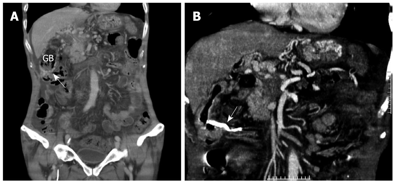
The patient continued to bleed despite endoscopic therapy. abdominal computed tomography (CT) was performed, which revealed portal vein tumor invasion with collateral circulation formation. Prominent varices were found around the gall bladder and cholecystojejunostomy (Figure 2A). No tumor invasion to the bowel was noted. Retrospective review of the endoscopic images revealed that the folds were mildly engorged with mild blue color, which suggested underlying varices.
After discussion, the patient agreed to endoscopic therapy with n-butyl-2-cyanoacrylate for these varices on the 12th d of hospitalization. Endoscopic ultrasound with miniprobe was used to confirm the presence of varices beneath the anastomotic ulcer, and injection therapy with n-butyl-2-cyanoacrylate was carried out smoothly (Figure 1C). A follow-up CT revealed successful obliteration of the collateral circulation (Figure 2B). The patient died of his disease 4 mo later and had no recurrent gastrointestinal bleeding during the intervening period.
Esophageal or gastric variceal bleeding is a common cause of severe gastrointestinal bleeding. Ectopic variceal bleeding from the duodenum[5-7], jejunum[8], ileum[9], and colon[10,11] have been reported in the literature as a diagnostic and therapeutic challenge to clinicians. This ectopic variceal bleeding usually results from portal hypertension, portal vein thrombosis, mesenteric vein thrombosis, chronic pancreatitis, adhesion after surgery, or inflammatory bowel disease[1,12]. Our reported case, suffering from cholecystojejunostomy variceal bleeding, is even rarer and such bleeding has been reported to be associated only with chronic pancreatitis[1-4] in the literature. This is probably because these cancer patients have a limited lifespan, dying before such varices can develop[1].
The diagnosis of ectopic varices is usually made after endoscopic examination: mesenteric venography[1,12], abdominal ultrasound[13], enteroclysis[3], or CT[14]. In our case, the diagnosis was difficult to make during the initial endoscopic examination because the varicose vein was not prominent and was masked by an overlying anastomotic ulcer. A careful review of the CT and endoscopic images in this case suggested the presence of varices in the cholecystojejunostomy and led us to approach the case with different endoscopic measures. Consequently, we suggest that endoscopic ultrasound is mandatory, as in our case, to confirm the presence of varices prior to endoscopic therapy. Alternatively, needle aspiration or injection of contrast may prove useful for diagnosis.
Unlike esophageal and gastric varices, the treatment options for ectopic varices have varied in the literature. Surgical resection[1] or radiological methods to decrease portal hypertension[15] have previously been reported. Both endoscopic band ligation[10] and sclerotherapy[15] have been successfully used to treat such ectopic varices. Endoscopic cyanoacrylate sclerotherapy was chosen in this patient with advanced cancer because it is minimally invasive and has previously been used to treat gastric varices endoscopically[16]. A follow-up CT demonstrated successful obliteration of the varices and thus, permanent hemostasis was achieved for our case.
In conclusion, we report a case of pancreatic cancer with bleeding from cholecystojejunal varices. The diagnosis was made by CT, endoscopy, and endoscopic ultrasound. Cyanoacrylate sclerotherapy was a successful method to achieve hemostasis in this case.
Peer reviewer: Richard A Kozarek, MD, Department of Gastroenterology, Virginia Mason Medical Center, 1100 Ninth Avenue, PO Box 900, Seattle, WA 98111-0900, United States
S- Editor Tian L L- Editor Logan S E- Editor Lin YP
| 1. | Carpenter S, Brown KA. Chronic complications after cholecystojejunostomy. Am J Gastroenterol. 1994;89:2073-2075. |
| 2. | Lein BC, McCombs PR. Bleeding varices of the small bowel as a complication of pancreatitis: case report and review of the literature. World J Surg. 1992;16:1147-1149; discussion 1150. |
| 3. | Miller JT Jr, De Odorico I, Marx MV. Cholecystojejunostomy varices demonstrated by enteroclysis. Abdom Imaging. 1997;22:474-476. |
| 4. | Getzlaff S, Benz CA, Schilling D, Riemann JF. Enteroscopic cyanoacrylate sclerotherapy of jejunal and gallbladder varices in a patient with portal hypertension. Endoscopy. 2001;33:462-464. |
| 5. | Omata F, Itoh T, Shibayama Y, Ide H, Takahashi H, Ueno F, Saubermann LJ, Matsuzaki S. Duodenal variceal bleeding treated with a combination of endoscopic ligation and sclerotherapy. Endoscopy. 1998;30:S62-S63. |
| 6. | Ota K, Shirai Z, Masuzaki T, Tanaka K, Higashihara H, Okazaki M, Arakawa M. Endoscopic injection sclerotherapy with n-butyl-2-cyanoacrylate for ruptured duodenal varices. J Gastroenterol. 1998;33:550-555. |
| 7. | Shiraishi M, Hiroyasu S, Higa T, Oshiro S, Muto Y. Successful management of ruptured duodenal varices by means of endoscopic variceal ligation: report of a case. Gastrointest Endosc. 1999;49:255-257. |
| 8. | Joo YE, Kim HS, Choi SK, Rew JS, Kim HR, Kim SJ. Massive gastrointestinal bleeding from jejunal varices. J Gastroenterol. 2000;35:775-778. |
| 9. | Misra SP, Dwivedi M, Misra V, Gupta M. Ileal varices and portal hypertensive ileopathy in patients with cirrhosis and portal hypertension. Gastrointest Endosc. 2004;60:778-783. |
| 10. | Soon MS, Yen HH, Soon A. Endoscopic band ligation for rectal variceal bleeding: serial colonoscopic images. Gastrointest Endosc. 2005;61:734-735. |
| 11. | Lopes LM, Ramada JM, Certo MG, Pereira PR, Soares JM, Ribeiro M, Areias J, Pinho C. Massive lower gastrointestinal bleeding from idiopathic ileocolonic varix: report of a case. Dis Colon Rectum. 2006;49:524-526. |
| 12. | Hodgson RS, Jackson JE, Taylor-Robinson SD, Walters JR. Superior mesenteric vein stenosis complicating Crohn's disease. Gut. 1999;45:459-462. |
| 13. | Ishida H, Konno K, Hamashima Y, Komatsuda T, Naganuma H, Asanuma Y, Ishida J, Masamune O. Small bowel varices: report of two cases. Abdom Imaging. 1998;23:354-357. |
| 14. | Smith TR. CT demonstration of ascending colon varices. Clin Imaging. 1994;18:4-6. |
| 15. | Hiraoka K, Kondo S, Ambo Y, Hirano S, Omi M, Okushiba S, Katoh H. Portal venous dilatation and stenting for bleeding jejunal varices: report of two cases. Surg Today. 2001;31:1008-1011. |
| 16. | Procaccini NJ, Al-Osaimi AM, Northup P, Argo C, Caldwell SH. Endoscopic cyanoacrylate versus transjugular intrahepatic portosystemic shunt for gastric variceal bleeding: a single-center U.S. analysis. Gastrointest Endosc. 2009;70:881-887. |


