Published online Mar 14, 2009. doi: 10.3748/wjg.15.1273
Revised: February 19, 2009
Accepted: February 26, 2009
Published online: March 14, 2009
Intraductal papillary mucinous neoplasm (IPMN) is an increasingly reported entity. Extensive pancreatic calcification is generally thought to be a sign of chronic pancreatitis, but it may occur simultaneously with IPMN leading to diagnostic difficulties. We report a case of a patient initially diagnosed with chronic calcifying pancreatitis who was later shown to have a malignant IPMN. This case illustrates potential pitfalls in the diagnosis of IPMN in the case of extensive pancreatic calcification as well as clues that may lead the clinician to suspecting the diagnosis. The possible mechanisms of the relation between pancreatic calcification and IPMN are also reviewed.
- Citation: Kalaitzakis E, Braden B, Trivedi P, Sharifi Y, Chapman R. Intraductal papillary mucinous neoplasm in chronic calcifying pancreatitis: Egg or hen? World J Gastroenterol 2009; 15(10): 1273-1275
- URL: https://www.wjgnet.com/1007-9327/full/v15/i10/1273.htm
- DOI: https://dx.doi.org/10.3748/wjg.15.1273
Intraductal papillary mucinous neoplasm (IPMN) is an increasingly reported entity, representing up to one third of all pancreatic cystic neoplasms[1]. IPMNs typically present with cystic dilatation of the pancreatic duct associated with mucin production and variable cellular atypia. Although extensive pancreatic calcification is generally considered to be a pathognomonic sign of chronic pancreatitis, it may also occur simultaneously with IPMN which may lead to diagnostic confusion[23]. We report a case of a patient who was initially diagnosed with chronic calcifying pancreatitis and was later shown to have an IPMN.
An 81-year-old man was referred to the gastro-enterology clinic due to weight loss, diarrhea, and anemia. He did not drink any alcohol and got quit of smoking pipe 50 years ago. Medical history was unremarkable apart from chronic obstructive pulmonary disease and type 2 diabetes mellitus diagnosed one year prior to presentation. Laboratory tests revealed mild normocytic normochromic anemia with 11.3 g/L hemoglobin (reference 13.0-17.0 g/L) but essentially normal white cell and platelet counts as well as normal electrolytes, creatinine, albumin, liver function tests, amylase, endomysial antibodies, plasma protein electrophoresis, and thyroid function tests.
Esophagogastroduodenoscopy was normal. A CT-colonography showed normal colonic appearances but there was evidence of a grossly atrophic calcific pancreas with a dilated pancreatic duct. No mass lesion was seen. The patient did not have a history of acute pancreatitis, abdominal radiotherapy or trauma to the epigastric region. His serum calcium and triglycerides were normal. Fecal elastase was < 15 &mgr;g/g of stool (reference > 500 &mgr;g/g of stools). Therefore , exocrine pancreatic insufficiency due to idiopathic chronic calcifying pancreatitis was diagnosed and pancreatic enzyme substitution was started.
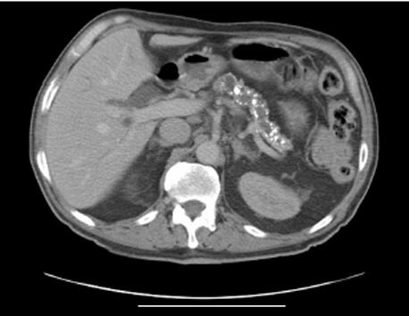
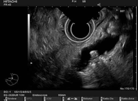
Despite some initial improvements, the patient reported further weight loss six months after initial presentation. Laboratory testing showed that his alkaline phosphatase was 2629 IU/L (reference 95-320 IU/L), bilirubin was 42 &mgr;mol/L (reference 3-17 &mgr;mol/L), and alanine aminotranferase was 347 IU/L (reference 10-45 IU/L). A pancreatic protocol CT demonstrated gross pancreatic atrophy and calcification, dilated pancreatic as well as intrahepatic and common bile ducts. No mass lesion was seen (Figure 1). Endoscopic ultrasound confirmed these findings and also showed an enlarged heterogeneous pancreatic head with dilated side branches of the pancreatic duct as well as an area of hypoechogeniccity, but did not show any distinct mass. The main pancreatic duct in the head was dilated with its diameter > 10 mm and with wall irregularities (Figure 2). Mucus extrusion from a protrubing gaping major papilla was observed. Endoscopic retrograde cholangiopancreatography was attempted in which opacification of the main pancreatic duct showed multiple filling defects suggestive of intraductal mucin. Common bile duct cannulation failed and biliary drainage was achieved by means of a percutaneous transhepatic cholangiography. Carcinoembryonic antigen (CEA) and carbohydrate antigen 19-9 (CA19-9) levels in mucin aspirated from the pancreatic duct were 24132 ng/mL and 60840 U/mL, respectively. On the basis of the imaging findings, the fact that mucus extrusion was observed from the papilla, and the high levels of CEA and CA 19-9 in pancreatic duct aspirates, a malignant IPMN was diagnosed. Due to his clinical condition, the patient was judged not to be a candidate for surgery and was offered palliative therapy.
Twelve to sixty percent of patients with IPMN have a history leading to a diagnosis of chronic pancreatitis[45] and roughly 2% of all diagnoses of chronic pancreatitis are associated with IPMN[5]. However, few reports of patients with concomitant calcyfing pancreatitis and IPMN are available[23]. In a cohort study of 473 patients suffering from chronic pancreatitis, only 6 were found to have IPMN during follow-up but none had calcifications[5]. Of the 16 cases of patients with IPMN and pancreatic calcification published thus far, 8 had diffuse pancreatic calcification which did not involve the tumor itself in most of the cases[23]. The pathogenesis of calcification in IPMN is unknown. Apart from the unlikely possibility that the two entities may occur coincidentally, it is possible that IPMN may be a complication of chronic calcifying pancreatitis, but no report of a patient with this condition developing IPMN in the course of the disease is available. It has also been proposed that IPMN may be responsible for pancreatic calcification due to chronic partial ductal obstruction by mucin plugs[23]. In any case, the concomitant presence of pancreatic cystic lesions, duct dilatation, and calcification should raise the suspicion of an IPMN. Particular care should be taken when diagnosing chronic pancreatitis in patients who do not present typical characteristics of the disease, namely age over 50 years, moderate alcohol intake, and non-smokers, which may be suggestive of IPMN[5]. In our case, the patient was 81 years of age and did not consume any alcohol as verified by the patient himself and his relatives.
Although CEA and CA 19-9 measurements are frequently performed during the diagnostic work-up of patients with pancreatic cystic lesions, it has been suggested that cyst fluid or pancreatic duct CEA or CA 19-9 is not helpful in the differentiation between benign and malignant IPMNs[6] but published data in the literature are not unanimous[7]. A CEA > 800 ng/mL has been reported to be helpful in differentiating mucinous cystadenocarcinomas from mucinous cystadenomas[8]. Although only 5/16 patients with concomitant calcifying pancreatitis and IPMN previously reported were shown to have a malignant lesion[23], in the present case the fact that both CEA and CA 19-9 in the pancreatic duct aspirate were extremely high was felt to be highly suggestive of malignancy. A main pancreatic duct > 6 mm in diameter and protrusion of the papilla of Vater are also considered signs predicting malignancy[9]. Thus, endoscopic ultrasound is not just a technique to obtain fluid from cystic pancreatic lesions for tumor marker analysis, but it also provides invaluable morphological information on mural pancreatic duct irregularities, presence of solid lesions or septa in cysts. Compared to other imaging methods, it achieves the highest detail resolution.
In summary, concomitant presence of pancreatic cystic lesions, duct dilatation, and calcification should raise the suspicion of an IPMN, especially when patients do not present typical characteristics of chronic pancreatitis, namely age over 50 years, moderate alcohol intake, and non-smokers. Inspection of the ampulla with a duodenoscope and aspiration of fluid from the pancreatic duct or endoscopic ultrasound-guided fine needle aspiration from a cystic pancreatic lesion for analysis of CEA and CA 19-9 may be useful in the diagnosis of such patients.
| 1. | Freeman HJ. Intraductal papillary mucinous neoplasms and other pancreatic cystic lesions. World J Gastroenterol. 2008;14:2977-2979. |
| 2. | Origuchi N, Kimura W, Muto T, Esaki Y. Pancreatic mucin-producing adenocarcinoma associated with a pancreatic stone: report of a case. Surg Today. 1998;28:1261-1265. |
| 3. | Zapiach M, Yadav D, Smyrk TC, Fletcher JG, Pearson RK, Clain JE, Farnell MB, Chari ST. Calcifying obstructive pancreatitis: a study of intraductal papillary mucinous neoplasm associated with pancreatic calcification. Clin Gastroenterol Hepatol. 2004;2:57-63. |
| 4. | Loftus EV Jr, Olivares-Pakzad BA, Batts KP, Adkins MC, Stephens DH, Sarr MG, DiMagno EP. Intraductal papillary-mucinous tumors of the pancreas: clinicopathologic features, outcome, and nomenclature. Members of the Pancreas Clinic, and Pancreatic Surgeons of Mayo Clinic. Gastroenterology. 1996;110:1909-1918. |
| 5. | Talamini G, Zamboni G, Salvia R, Capelli P, Sartori N, Casetti L, Bovo P, Vaona B, Falconi M, Bassi C. Intraductal papillary mucinous neoplasms and chronic pancreatitis. Pancreatology. 2006;6:626-634. |
| 6. | Pais SA, Attasaranya S, Leblanc JK, Sherman S, Schmidt CM, DeWitt J. Role of endoscopic ultrasound in the diagnosis of intraductal papillary mucinous neoplasms: correlation with surgical histopathology. Clin Gastroenterol Hepatol. 2007;5:489-495. |
| 7. | Kawai M, Uchiyama K, Tani M, Onishi H, Kinoshita H, Ueno M, Hama T, Yamaue H. Clinicopathological features of malignant intraductal papillary mucinous tumors of the pancreas: the differential diagnosis from benign entities. Arch Surg. 2004;139:188-192. |
| 8. | van der Waaij LA, van Dullemen HM, Porte RJ. Cyst fluid analysis in the differential diagnosis of pancreatic cystic lesions: a pooled analysis. Gastrointest Endosc. 2005;62:383-389. |


