Published online Sep 21, 2007. doi: 10.3748/wjg.v13.i35.4781
Revised: June 1, 2007
Accepted: June 9, 2007
Published online: September 21, 2007
Extragonadal germ cell tumors are rare. The most common sites for EGGCTs are in midline locations such as the mediastinum, retroperitoneum and pineal gland. These tumors rarely present in the stomach. We describe here a case where a middle aged man presented with typical symptoms of gastric cancer. After extensive workup, which included blood work, CT abdomen scan, upper endoscopy, and endoscopic ultrasound, the patient was diagnosed with gastric cancer. However, due to very high blood levels of alpha-fetoprotein, the specimen was sent for special histochemical staining, which demonstrated that the tumor had features of both adenocarcinoma and endodermal sinus tumor. This is a very aggressive tumor with a very poor prognosis.
- Citation: Singh M, Arya M, Anand S, Sandar N. Gastric adenocarcinoma with features of endodermal sinus tumor. World J Gastroenterol 2007; 13(35): 4781-4783
- URL: https://www.wjgnet.com/1007-9327/full/v13/i35/4781.htm
- DOI: https://dx.doi.org/10.3748/wjg.v13.i35.4781
Extragonadal germ cell tumors are rare, accounting for only 1% to 4% of all germ cell tumors. Ninety-five percent of all testicular tumors are germ cell tumors. These tumors originate in the sperm forming cells in the testicles (the male gonads) or egg producing cells in the ovary (female gonads). On rare occasions, however, germ cell tumors develop elsewhere in the body without any evidence of cancer in the testes. When this happens, they are referred to as extragonadal germ cell tumors (EGGCTs) (from the Testicular Cancer Resource Center)[1].
At approximately 4 to 6 wk of embryonic development, the germ cells migrate into the embryo where they populate the developing testes or ovaries. If these cells miss their destination, they are likely to come to rest in one of a number of midline sites in the body. Extragonadal tumors arise when these cells become cancerous.
EGGCTs can be either benign or malignant and the malignant tumors can be either seminoma or non-seminoma. Extragonadal tumors are much more common in females. Malignant extragonadal tumors are much more common in men.
Extragonadal tumors can arise anywhere in the body. But, the majority are found in 3 common sites: anterior mediastinum, the retroperitoneum, and pineal gland in the brain. EGGCTs are aggressive and are usually seen in young adults. The treatment and prognosis of the disease depends on a variety of factors including the type of cancer, the tumor location, and the size of the tumor.
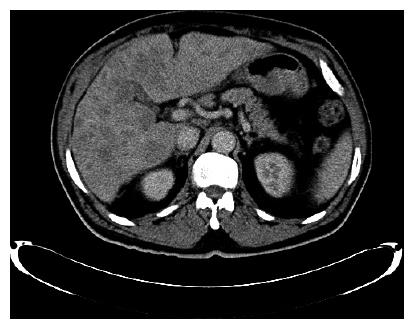
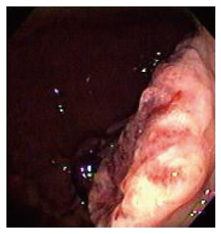
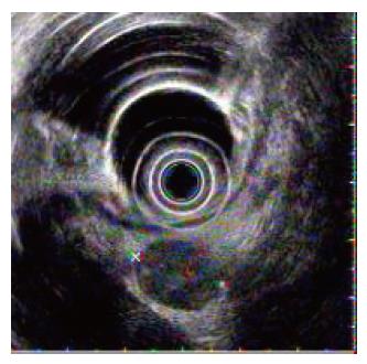
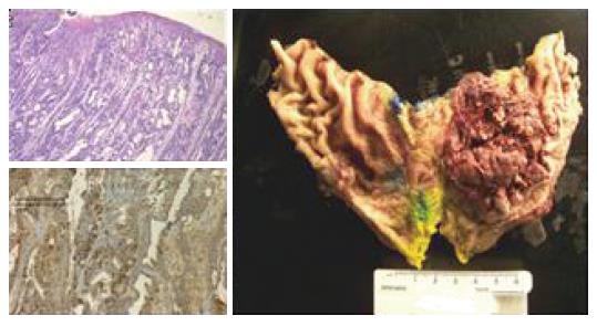
A 67-year-old Hispanic male presented to our institution with a complaint of epigastric pain radiating to the right upper quadrant, 8/10 in intensity, and it worsened with eating for 2 to 3 mo. The patient also complained of a weight loss of 10 pounds over the past month and vomiting for 2 d. He denied melena, hematochezia, and hematemesis. Past medical history was only significant for hyperlipidemia. The patient smoked 1 pack of cigarettes for 40 years, but denied use of alcohol. Family history was only significant for diabetes and hypertension. On physical examination, the patient felt a slight pain on deep palpation in the epigastrium with voluntary guarding. The remainder of his physical examination, including genital examination, was normal. Initial blood work obtained in the emergency room showed iron deficiency anemia, and elevated AST, ALT, and alkaline phosphatase. Tumor markers were sent and his serum alpha-fetoprotein was very high (4178; normal 0-1.8). Beta-HCG was normal. A CT scan of the abdomen showed gastric wall thickening and several 2 cm lymph nodes in mesenteric fat inferior to the stomach and liver extensively replaced with metastatic disease (Figure 1). An upper endoscopy, and endoscopic ultrasound were obtained (Figure 2 and Figure 3). Endoscopy showed a large 8-10 cm exophytic-ulcerating lesion along the greater curvature, extending from mid body to the antrum. A tissue biopsy was obtained. EUS demonstrated a mass extending through all layers of the stomach. Tissue biopsy obtained via EGD demonstrated invasive adenocarcinoma, which was poorly differentiated, and there were no H Pylori organisms. In view of the patient’s very high serum alpha fetoprotein, a mixed component of extragonadal germ cell and adenocarcinoma was suspected, and the biopsy specimen was sent for special immunohistochemical staining (Figure 4). The immunohistochemical stains for alpha-fetoprotein strongly reacted with the tumor cells. Synaptophysin was focally positive and chromogranin was negative. These findings support the diagnosis of gastric adenocarcinoma, which was moderate to poorly differentiated, with features of endodermal sinus tumor and endocrine type cells.
The patient underwent exploratory laparotomy with partial gastric resection. The patient also received platinum based chemotherapy with Platinol and Taxol. He had a very eventful 2 mo of hospital course with multiple infections, upper gastrointestinal bleeding and eventual respiratory failure and death.
Extragonadal germ cell tumors are rare, representing 1% to 4% of all germ cell tumors. Seminomas account for 30% to 40% of these tumors, and non-seminomas account for 60% to 70%. The most common site of EGGCTs is the mediastinum (50%-70%) followed by the retro peritoneum (30%-40%), the pineal gland (5%), and the sacrococcygeal area (less than 5%). In children, benign and malignant EGGCTs occur equally in males and females. In adults, only benign EGGCTs (teratomas) occur at equal frequency in both sexes; more than 90% of malignant EGGCTs occur in males[2].
Symptoms vary depending on the site and the size of the tumor. A complete physical examination is required. Workup of EGGCTs begins with lab studies with the tumor markers alpha-fetoprotein and beta-HCG. These tumor markers provide diagnostic, staging and prognostic information. Pure seminomas and pure choriocarcinomas do not produce AFP. A CT scan of the chest, abdomen, and pelvis should be obtained. Treatment consists of surgery and chemotherapy. A classification system has been developed by the International Germ Cell Collaborative Group (IGCCG). This system categorizes tumors on the basis of histologic type, localization of metastases, and initial levels of serum AFP, beta-HCG, and LDH. Our patient who had very high levels of AFP and liver metastases expired within two months of the diagnosis[3].
As mentioned earlier, these tumors generally present at an early age. However, few cases of EGGCTs in the elderly have been described in the literature[4]. A case of yolk sac tumor with adenocarcinoma has been described in a 56-year-old female[5]. On even rarer occasions, a gastric adenocarcinoma can present with a combination of these tumors. A case with such a tumor has been described in a Korean pathology journal where gastric adenocarcinoma was mixed with gastric choriocarcinoma and endodermal sinus tumor[5].
S- Editor Liu Y L- Editor Knapp E E- Editor Lu W
| 1. | The testicular cancer resource center. 1997-2006. Available from: http//www.acor.org/tcrc/egc.html. |
| 2. | Makhoul I. The Oregon Clinic. 2004;1-9. |
| 3. | Motoyama T, Saito K, Iwafuchi M, Watanabe H. Endodermal sinus tumor of the stomach. Acta Pathol Jpn. 1985;35:497-505. [PubMed] |
| 4. | Suzuki T, Kimura N, Shizawa S, Yabuki N, Yamaki T, Sasano H, Nagura H. Yolk sac tumor of the stomach with an adenocarcinomatous component: a case report with immunohistochemical analysis. Pathol Int. 1999;49:557-562. [RCA] [DOI] [Full Text] [Cited by in Crossref: 15] [Cited by in RCA: 19] [Article Influence: 0.7] [Reference Citation Analysis (0)] |
| 5. | Kim EK, Hong EK, Lee KS, Lee JD. Gastric choriocarcinoma and endodermal sinus tumor in collision tumor. Korean J Pathol. 1992;26:405-410. |


