Published online Feb 7, 2006. doi: 10.3748/wjg.v12.i5.825
Revised: June 19, 2005
Accepted: July 28, 2005
Published online: February 7, 2006
This report describes an extremely rare adult case of an omphalomesenteric cyst resected by laparoscopic-assisted surgery. A 29-years-old Japanese man was referred and admitted to Kyushu University Hospital because of an abdominal mass and an elevated serum CEA (carcinoembryonic antigen) level (21.3 ng/mL) in August 2001. Abdominal CT and US demonstrated a cystic mass with septum and calcification. Laparoscopy showed a large mass to be attached to his abdominal wall, measuring 110 mm x 70 mm x 50 mm and filled with mucus. The mass was resected by laparoscopic-assisted surgery. The histological findings of its wall showed fibromuscular tissue, adipose tissue, calcification, and an intestinal structure. It was finally diagnosed to be an omphalomesenteric cyst.
- Citation: Sawada F, Yoshimura R, Ito K, Nakamura K, Nawata H, Mizumoto K, Shimizu S, Inoue T, Yao T, Tsuneyoshi M, Kondo A, Harada N. Adult case of an omphalomesenteric cyst resected by laparoscopic-assisted surgery. World J Gastroenterol 2006; 12(5): 825-827
- URL: https://www.wjgnet.com/1007-9327/full/v12/i5/825.htm
- DOI: https://dx.doi.org/10.3748/wjg.v12.i5.825
The omphalomesenteric duct remnant is one of the rare congenital anomalies associated with the primitive yolk stalk. Most omphalomesenteric duct remnants tend to be Meckel’s diverticulum, however, the occurrence of an omphalomesenteric cyst is very rare. An omphalomesenteric duct remnant induces several symptoms, such as intestinal obstruction, abdominal pain, melena, and umbilical hernia. These symptoms tend to occur most frequently during the childhood years. In this report, we have described an extremely rare adult case of omphalomesenteric cyst resected by laparoscopic-assisted surgery.
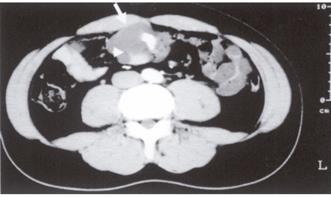
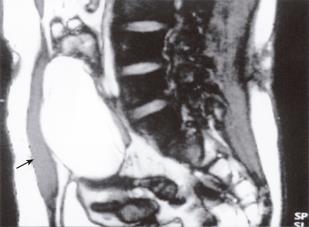
A 29-year-old male patient visited Aso Iizuka Hospital with the complaint of an abdominal mass of one month duration in July 2001. The laboratory data revealed an elevated CEA level (21.3 ng/mL). Abdominal CT showed a cystic mass, suspected of being a mesenteric cyst. In August 2001, he was referred and admitted to Kyushu University Hospital for further management. Physical examinations showed no abnormalities except for a fist-sized, elastic soft mass in his umbilical region. The laboratory data demonstrated elevations in the ALT (32I U/L), CEA (7.9 ng/mL, normal range <3.2 ng/mL), and CA19-9 (62.6 U/mL, normal range <37.0 U/mL) levels. However, the serum CA125 and alpha fetoprotein (AFP) levels were both within the normal limits. Abdominal US and CT showed a cystic mass with septum and calcification, measuring 100 mm x 80 mm x50 mm in size (Figure 1). MRI (T2-WI) showed a cystic mass with septum, which was suspected to be a mesenteric cyst (Figure 2). A barium small-bowel enema showed no abnormality except for extraductal oppression. Angiography showed an avascular area in the mass. Our preoperative diagnosis was mesenteric cyst.
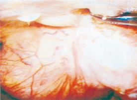
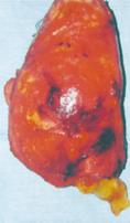
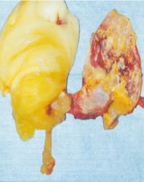
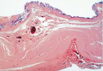
Laparoscopic-assisted surgery was performed for the purpose of making a precise diagnosis and resection, because the possibility of a CEA-producing malignant tumor first had to be ruled out. A laparoscope was inserted through a 1 cm-trocar site at the median upper abdomen. Laparoscopy showed the mass to be attached to his abdominal wall at the median hypogastrium (Figure 3), and two ligaments, considered to be remnants of the umbilical arteries, at its attached portion. The mass had no communication with his small bowel. There was no evidence of a persistent urachus. Next, an additional two ports were made with one at the right upper abdomen and another at the right lateral abdomen. The mass was then resected from his abdominal wall using laparoscopic scissors and electrocautery. The mass was removed through a 5-cm median incision made at his lower abdomen. The mass measured 110 mm x 70 mm x 50 mm in size (Figure 4), and weighed 160 g. This cystic mass was filled with mucus (Figure 5). The histological findings demonstrated the cystic wall to be composed of fibromuscular tissue, adipose tissue, focal calcification and an intestinal type mucosa with goblet cells, accompanied by ulceration with a regenerative epithelium (Figure 6). The histological findings showed no evidence of malignancy. It was finally diagnosed to be an omphalomesenteric cyst. Six months after the operation, the serum CEA and CA19-9 levels both became normal (0.7 ng/mL and 12.46 U/mL, respectively).
Omphalomesenteric duct remnants (vitelline duct anomalies) have been reported to be congenital anomalies associated with the primitive yolk stalk. The omphalomesenteric duct is the embryonic structure connecting the primary yolk sac to the embryonic midgut that normally closes off spontaneously at about 5-9 wk of gestation[1]. The failure of such closure may result in various lesions occurring in the omphalomesenteric duct remnants, such as Meckel’s diverticulum, complete umbilical enteric fistula, incomplete umbilical sinus, omphalomesenteric cyst, or umbilical mucosal polyp[2]. Most of omphalomesenteric duct remnants (67%) tend to develop into Meckel’s diverticulum, however, omphalomesenteric cyst is also rarely observed[3]. Vane et al[1] reported only three cases of omphalomesenteric cyst in 217 children with omphalomesenteric duct remnants. The etiology of omphalomesenteric cyst is a persistent lesion of the intermediate duct with closure at both ends, leaving a cyst which can attach itself to the umbilicus, bowel, or both[4].
Omphalomesenteric duct remnants may persist in approximately 2% of infants[4,5]. Vane et al [1] reported the male-female ratio in a 217 remnant series to be 2:1. Yamada et al [6] reported the male-female ratio to be 2.8:1 in a Japanese series of 65 cases. The male-female ratio of omphalomesenteric cyst was reported to be 4:1 which correlates with the previously reported male predominance in other omphalomesenteric duct anomalies[7].
Common symptoms of omphalomesenteric duct remnants include abdominal pain, rectal bleeding, intestinal obstruction, umbilical drainage, and umbilical hernia[3], and all these symptoms appear to be age-dependent. In addition, most of the symptoms usually appear before the age of 4 years[1]. Eighty-five percent of infants younger than 1 month and 77% of children aged 1 month to 2 years had a symptomatic presentation[1]. It was also reported that 40% of the children with this anomaly had symptomatic lesions, while this anomaly is usually asymptomatic in adults[1]. Adult cases of omphalomesenteric cyst are extremely rare.
A surgical resection is necessary for symptomatic omphalomesenteric duct remnants, but not necessary for asymptomatic omphalomesenteric duct remnants[8,9]. In our case, although asymptomatic omphalomesenteric cyst was incidentally found, it was removed because the possibility of a CEA-producing malignant tumor and small-bowel obstruction had to be ruled out[1]. The elevated tumor markers (CEA, CA19-9) all returned to normal levels after the resection. However, its histological findings showed no evidence of malignancy. The etiology of such elevated tumor markers remains to be elucidated.
In our case, the omphalomesenteric cyst was resected safely by laparoscopic-assisted surgery. Recent advances in diagnostic modalities have resulted in an increased incidental discovery of asymptomatic congenital anomalies. In such cases, the use of laparoscopic surgery is considered to be an effective, safe and less invasive treatment.
S- Editor Kumar M and Guo SY L- Editor Elsevier HK E- Editor Li HY
| 1. | Vane DW, West KW, Grosfeld JL. Vitelline duct anomalies. Experience with 217 childhood cases. Arch Surg. 1987;122:542-547. [RCA] [PubMed] [DOI] [Full Text] [Cited by in Crossref: 113] [Cited by in RCA: 102] [Article Influence: 2.7] [Reference Citation Analysis (0)] |
| 2. | Moore TC. Omphalomesenteric duct malformations. Semin Pediatr Surg. 1996;5:116-123. [PubMed] |
| 3. | Lassen PM, Harris MJ, Kearse WS, Argueso LR. Laparoscopic management of incidentally noted omphalomesenteric duct remnant. J Endourol. 1994;8:49-51. [RCA] [PubMed] [DOI] [Full Text] [Cited by in Crossref: 12] [Cited by in RCA: 11] [Article Influence: 0.4] [Reference Citation Analysis (0)] |
| 4. | Quarantillo EP. Cyst of the omphalomesenteric duct presenting as an acute abdominal condition. Am J Surg. 1967;114:465-466. [RCA] [PubMed] [DOI] [Full Text] [Cited by in Crossref: 4] [Cited by in RCA: 4] [Article Influence: 0.1] [Reference Citation Analysis (0)] |
| 5. | Shaw A. Disorders of the umbilicus. Pediatric Surgery. 4th ed Chicago: YearBook Medical Publisher Inc 1986; 731-739. |
| 6. | Hawkins R, Hass WK, Ransohoff J. Advantages of 2-14C glucose for regional cerebral glucose utilization. Acta Neurol Scand Suppl. 1977;64:436-437. [PubMed] |
| 7. | Heifetz SA, Rueda-Pedraza ME. Omphalomesenteric duct cysts of the umbilical cord. Pediatr Pathol. 1983;1:325-335. [RCA] [PubMed] [DOI] [Full Text] [Cited by in Crossref: 32] [Cited by in RCA: 33] [Article Influence: 0.8] [Reference Citation Analysis (0)] |
| 8. | Hinson RM, Biswas A, Mizelle KM, Tunnessen WW. Picture of the month. Persistent omphalomesenteric duct. Arch Pediatr Adolesc Med. 1997;151:1161-1162. [RCA] [PubMed] [DOI] [Full Text] [Cited by in Crossref: 9] [Cited by in RCA: 9] [Article Influence: 0.3] [Reference Citation Analysis (0)] |
| 9. | Townsend CM Jr, Thompson JC. Small intestine. Principles of Surgery. 5th ed. New York: McGraw-Hill 1989; 1212. |


