Copyright
©2006 Baishideng Publishing Group Co.
World J Gastroenterol. Feb 7, 2006; 12(5): 825-827
Published online Feb 7, 2006. doi: 10.3748/wjg.v12.i5.825
Published online Feb 7, 2006. doi: 10.3748/wjg.v12.i5.825
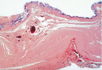
Figure 6 Histological features showing cystic wall to be composed of fibromuscular tissue, adipose tissue, focal calcification, and an intestinal structure, lined by an intestinal type mucosa with goblet cells, accompanied by ulceration with a regenerative epithelium.
- Citation: Sawada F, Yoshimura R, Ito K, Nakamura K, Nawata H, Mizumoto K, Shimizu S, Inoue T, Yao T, Tsuneyoshi M, Kondo A, Harada N. Adult case of an omphalomesenteric cyst resected by laparoscopic-assisted surgery. World J Gastroenterol 2006; 12(5): 825-827
- URL: https://www.wjgnet.com/1007-9327/full/v12/i5/825.htm
- DOI: https://dx.doi.org/10.3748/wjg.v12.i5.825


