Published online Jan 21, 2005. doi: 10.3748/wjg.v11.i3.396
Revised: June 4, 2004
Accepted: July 17, 2004
Published online: January 21, 2005
AIM: To employ pulse wave Doppler ultrasonography to evaluate the changes in portal blood flow velocity in patients with chronic hepatitis C (CHC) receiving interferon (IFN) treatment.
METHODS: The subjects in this study were 14 patients (13 men and 1 woman) with CHC who received IFN treatment. Portal blood flow velocity was measured in the vessels at the porta hepatis at four time points: before IFN administration (pre-IFN), 2 wk after the start of administration (wk 2), 24 wk after the start of administration (wk 24, i.e., the end of IFN administration), and 24 wk after the end of administration (wk 48).
RESULTS: The patients with CHC in whom IFN treatment resulted in complete elimination or effective elimination of viruses showed a significant increase in portal blood flow velocity at the end of IFN treatment compared with that before IFN treatment. In contrast, when IFN was ineffective, no significant increase in portal blood flow velocity was observed at wk 24 or 48 compared with the pre-IFN value. In addition, the patients with CHC in whom IFN was ineffective showed significantly lower portal blood flow velocity values than control subjects at all measurement time points.
CONCLUSION: Pulse wave Doppler ultrasonography is a noninvasive and easily performed method for evaluating the effects of IFN treatment in patients with CHC. This technique is useful for measuring portal blood flow velocity before and 24 wk after IFN administration in order to evaluate the changes over time, thus assessing the effectiveness of IFN treatment.
- Citation: Nakanishi S, Shiraki K, Yamamoto K, Koyama M, Kimura N, Nakano T. Hemodynamics in the portal vein evaluated by pulse wave Doppler ultrasonography in patients with chronic hepatitis C treated with interferon. World J Gastroenterol 2005; 11(3): 396-399
- URL: https://www.wjgnet.com/1007-9327/full/v11/i3/396.htm
- DOI: https://dx.doi.org/10.3748/wjg.v11.i3.396
A number of studies have reported that interferon (IFN) is effective for the treatment of chronic hepatitis C (CHC) and have demonstrated improvements in hepatic function biochemically, histologically, and virologically[1-4]. In addition, IFN has been found to improve the prognosis of patients because it has an anticarcinogenic effect on the progression of hepatic cirrhosis[5,6].
In assessing the effectiveness of IFN treatment on patients with CHC, there are unequivocal indices in clinical laboratory tests, such as hepatitis C virus RNA (HCV-RNA) and alanine aminotransferase (ALT) levels, which can also be expected to serve as indicators of the complete elimination of HCV-RNA by IFN (sustained viral response: SVR). However, IFN is not effective in all patients with CHC. The effectiveness of IFN treatment has been reported to vary depending on a number of background factors such as the HCV-RNA value before IFN administration, the HCV genotype, and the histopathological stage[7,8]. These factors can be evaluated before the start of IFN administration in order to predict the effectiveness of IFN treatment.
In the meantime, ultrasonography has been widely recognized as a noninvasive, easy-to-perform diagnostic imaging modality that is indispensable for assessing the condition of the liver in patients with CHC. In addition, pulse wave Doppler ultrasonography, which is based on the Doppler effect, has been widely employed in clinical practice. However, the only study that has described the use of pulse wave Doppler ultrasonography for assessing the effectiveness of IFN treatment was the study by Walsh et al[14] in 1998, which reported the 12-wk follow-up results after treatment with interferon-alpha in patients with CHC.
The objectives of the present study were to employ pulse wave Doppler ultrasonography to measure the portal blood flow velocity before and after IFN administration in patients with CHC, to identify the correlations between IFN treatment effectiveness and findings of portal blood flow velocity analysis, and to assess the usefulness of portal blood flow velocity analysis in evaluating the effectiveness of IFN treatment.
The subjects in this study were 14 patients with CHC who received IFN treatment at Matsuzaka Chuo General Hospital from March 2002 to October 2003 (13 men and 1 woman; age range, 35 to 65 years; mean age, 52.9 years). The study also included 15 control subjects (CS group) (10 men and 5 women; age range, 35-55 years; mean age, 48.6 years). All of the patients with CHC underwent liver biopsy before the start of IFN administration, and the tissue type was identified by histopathological examination. The HCV genotype was also determined (Table 1).
| CHC | CS | |
| n | 14 | 15 |
| Sex | Male: 13, Female: 1 | Male: 10, Female: 5 |
| Age (yr) | 35-65 (52.9) | 30-55 (48.6) |
| Genotype | 1b:8, 2a:5, 2b:1 | |
| Activity | A1:11, A2:3 | |
| Fibrosis | F1:9, F2:5 |
Two IFN administration protocols were employed. One administration protocol was the combination of IFNα 2b (6 million units) and ribavirin (600 mg/d in patients weighing less than 60 kg and 800 mg/d in patients weighing more than 60 kg). The other administration protocol was IFNαcon-1 (18 million units). IFN was administered continuously for the first 2 wk and 3 times per week from wk 3 to wk 24. Administration was stopped at wk 24. In addition, the effectiveness of treatment was evaluated 24 wk after the end of IFN administration (wk 48). In the assessment of treatment effectiveness, the criteria for complete elimination (IFN-SVR) were a negative HCV-RNA result at wk 48 as well as an ALT value within the normal range. The criteria for effective elimination (IFN-BR) were a positive HCV-RNA result at wk 48 and an ALT value within the normal range. If the above criteria were not met, the result was judged to be no response (IFN-NR).
Portal blood flow velocity was evaluated at four time points: before IFN administration (pre-IFN), 2 wk after the start of IFN administration (wk 2), at the end of IFN administration (wk 24), and 24 wk after the end of IFN administration (wk 48).
ALT (IU/L) and HCV-RNA (kIU/mL) values were measured at the specified time points, and portal blood flow velocity analysis was performed. Portal blood flow velocity was measured using a diagnostic ultrasound system (Toshiba SSA-370, Power Vision 6000) at the specified time points to obtain the mean velocity (PVV mean cm/s) in the vessels at the porta hepatis. A convex-type transducer (center frequency, 3.5 MHz) was employed to examine the major vessels in the porta hepatis by right intercostal scanning or hypochondrial oblique scanning.
For portal blood flow velocity analysis, the measurement sample volume was set slightly smaller than the lumen of the target vessel in order to minimize noise. In addition, the angle of incidence of the ultrasound beam relative to the vessel was set to not more than 60o. Subsequently, a specified period of the Doppler signals obtained by pulse wave Doppler ultrasonography (PDUS) was extracted, and the Doppler shift was calculated at a high speed by fast Fourier transform (FFT). The shift frequency spectrum obtained was then used to generate images, and the waveforms were displayed. The waveforms were traced over two cardiac cycles, and the mean velocities of these waveforms were automatically calculated. This procedure was repeated five times and the mean value was used.
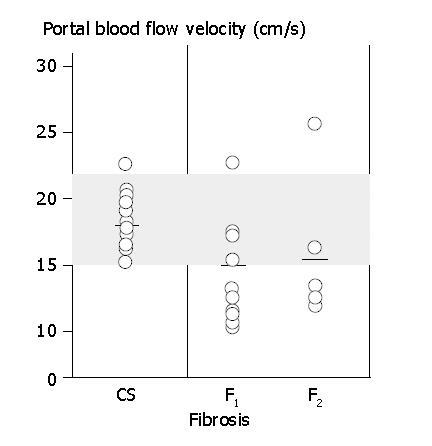
The portal blood flow velocity (PVV) was 18.3±3.2 cm/s (mean±SD) in the healthy adult volunteers (CS group).
The effectiveness of IFN treatment was evaluated based on the HCV-RNA values and HCV genotype. Of the 8 patients with genotype 1b, 4 were judged to have SVR (50%), 2 were judged to have BR (25%), and 2 were judged to have NR (25%). In addition, 3 of the 4 patients who were judged to have SVR had HCV-RNA levels less than 100 kIU/mL. Of the 5 patients with genotype 2a, 3 were judged to have SVR (60%) and 2 were judged to have NR (40%). One patient with genotype 2b had a HCV-RNA level greater than 1000 kIU/mL but was judged to have SVR.
With regard to the assessment of disease activity in patients with chronic hepatitis who received IFN treatment, the effectiveness of IFN treatment in 11 patients classified as A1 was judged to have SVR in 6 patients (55%), BR in 2 patients (18%), and NR in 3 patients (27%). Treatment effectiveness in 3 patients classified as A2 was judged to have SVR in 2 patients (66%) and NR in 1 patient (34%). With regard to the changes in portal blood flow velocity, no significant differences were observed between the CS group and the active disease A1 and A2 groups. When IFN was administered to CHC patients with different disease activity classifications (A1 and A2), no significant differences were observed in either group with regard to portal blood flow velocity before and after IFN administration, indicating comparable levels of treatment effectiveness in both A1 and A2 groups.
Next, the evaluation based on the degree of fibrosis showed that the treatment effectiveness was SVR in 6 (66%) of the 9 patients classified as F1, and in 2 (40%) of the 5 patients classified as F2. Assessment of changes in portal blood flow velocity showed no significant differences between F1 and F2 groups. In addition, no significant differences were observed between F1, F2, and CS groups (Figure 1). However, the portal blood flow velocity at wk 24 was significantly increased (P<0.05) compared with that before IFN treatment (F1: 14.2±4.3 cm/s→16.6±3.5 cm/s, F2: 15.4±6.1 cm/s→16.8±cm/s).
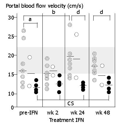
The changes in portal blood flow velocity at various time points of IFN treatment were also evaluated. The portal blood flow velocity in patients judged to have NR was 11.5±1.0 cm/s before IFN administration, significantly lower than that in the CS group (P<0.001). Furthermore, the portal blood flow velocities in patients judged to have NR at wk 2, wk 24, and wk 48 were 12.9±1.3 cm/s (P<0.001), 13.2±0.4 cm/s (P<0.001), and 14.6±1.6 cm/s (P<0.05) respectively all of which were significantly lower than the values in the CS group. Portal blood flow velocities in patients judged to have SVR and BR before IFN treatment and at wk 2, wk 24, and wk 48 were 16.9±6.0 cm/s, 14.9±2.5 cm/s, 18.7±4.0 cm/s, and 16.9±2.4 cm/s respectively. The portal blood flow velocity at wk 24 was significantly higher than the pre-IFN velocity (P<0.01). Portal blood flow velocity was also compared between the SVR, BR, and NR groups. The patients in the NR group showed significantly lower values before IFN treatment (P<0.05), at wk 24 (P<0.001), and at wk 48 (P<0.001) (Figure 2).
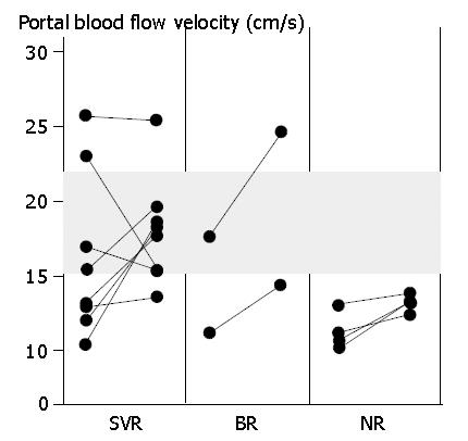
The increases or decreases in portal blood flow velocity (i.e., a change in velocity of 20% or more) were compared between pre-IFN values and those obtained at wk 24. In a total of 14 patients, the portal blood flow velocity at wk 24 was increased in 7 patients (50%), unchanged in 6 patients (43%), and decreased in 1 patient (7%), compared with the pre-IFN velocity. Comparisons were also made based on the differences in IFN treatment effectiveness. In the SVR group (8 patients), the velocity was increased in 4 patients (50%), unchanged in 3 patients (38%), and decreased in 1 patient (13%). An increase was therefore the most common finding in this group. In the BR group (2 patients), an increase was seen in both patients. However, in the NR group (4 patients), the velocity was increased in 1 patient (25%) and unchanged in 3 patients (75%), indicating that the majority of patients in this group had no change. In summary, portal blood flow velocity was increased in 7 of the 14 patients, and 6 of these 7 patients (86%) were judged to have SVR or BR (Figure 3).
A number of reports have analyzed chronic liver diseases in relation to portal hemodynamics as assessed by pulse wave Doppler ultrasonography[8-12]. Ramazan et al[12] examined 75 healthy subjects and reported that the blood flow velocity in the main portal vessel was 17.3±9.5 cm/s.
The PVV in the CS group in the present study was 18.3±3.2 cm/s, which is in good agreement with the value reported by Ramazan et al[12]. No significant differences in portal blood flow velocity were seen between the CS group and the 14 patients with CHC examined in this study. However, the pre-IFN portal blood flow velocity in the IFN-NR group was significantly lower than that in the CS group. The follow-up results after administration also showed that the velocity was significantly lower in the IFN-NR group than in the CS group. A number of studies have reported no significant differences in portal blood flow velocity between patients with chronic hepatic diseases and healthy subjects[8-13]. In the present study, however, although the overall portal blood flow velocity in the CHC group was not significantly different from that in the CS group, when the NR group alone was considered, the portal blood flow velocity was significantly lower than that in the CS group.
Walsh et al[14] employed pulse wave Doppler ultrasonography in the examination of 39 patients with CHC who received a 12-wk IFN-alpha treatment, and reported the Doppler perfusion index (DPI) (calculated as the ratio of hepatic artery flow to total hepatic flow) and the congestive index of the portal vein (area/velocity). According to their report, these indices did not change following a 12-wk IFN-alpha treatment. In the present study, the IFN-NR group showed no significant changes in portal blood flow velocity at 24 wk after IFN treatment, which is similar to the findings of Walsh et al[14] Nevertheless, the portal blood flow velocity in the SVR and BR groups was significantly increased at 24 wk after IFN treatment, as compared with that before treatment.
In the present study, the effectiveness of IFN treatment was also evaluated in relation to the histopathological findings obtained by liver biopsy. No significant differences were observed between two indices of the degree of liver fibrosis (i.e., between the F1 group with fibrous dilatation in the portal area and the F2 group with bridging fibrosis). Di Bisceglie et al[2] and Omata et al[15] performed histological assessments of IFN treatment in patients with chronic hepatitis C and non-A, non-B chronic hepatitis before and after IFN treatment. The results of these studies demonstrated clear improvements in periportal necrosis, focal necrosis, and portal inflammation, but not in fibrosis. It is particularly important to note the clear evidence of improvement in periportal necrosis and portal inflammation. In the IFN-SVR group, periportal necrosis and portal inflammation associated with increased portal resistance and reduced portal blood flow velocity, occurred before IFN treatment. These pathologic changes were improved by IFN treatment, presumably resulting in a reduction in portal resistance and an increase in portal blood flow velocity.
In summary, the present study was conducted to assess the usefulness of portal blood flow velocity analysis using pulse wave Doppler ultrasonography (PDUS) in evaluating the effectiveness of IFN treatment. The results of this study demonstrated that patients judged to have IFN-SVR and IFN-BR showed a significantly higher portal blood flow velocity at the end of IFN treatment than before IFN treatment. In addition, no response to IFN was suggested when the portal blood flow velocity at the end of IFN treatment was not significantly higher than the pre-IFN value. In the assessment of the effects of IFN treatment in patients with chronic hepatitis C, pulse wave Doppler ultrasonography permits the portal blood flow velocity to be easily measured and is therefore considered to be a clinically useful method for evaluating treatment effectiveness.
Edited by Wang XL
| 1. | Davis GL, Balart LA, Schiff ER, Lindsay K, Bodenheimer HC, Perrillo RP, Carey W, Jacobson IM, Payne J, Dienstag JL. Treatment of chronic hepatitis C with recombinant interferon alfa. A multicenter randomized, controlled trial. Hepatitis Interventional Therapy Group. N Engl J Med. 1989;321:1501-1506. [RCA] [PubMed] [DOI] [Full Text] [Cited by in Crossref: 1213] [Cited by in RCA: 1130] [Article Influence: 31.4] [Reference Citation Analysis (0)] |
| 2. | Di Bisceglie AM, Martin P, Kassianides C, Lisker-Melman M, Murray L, Waggoner J, Goodman Z, Banks SM, Hoofnagle JH. Recombinant interferon alfa therapy for chronic hepatitis C. A randomized, double-blind, placebo-controlled trial. N Engl J Med. 1989;321:1506-1510. [RCA] [PubMed] [DOI] [Full Text] [Cited by in Crossref: 900] [Cited by in RCA: 863] [Article Influence: 24.0] [Reference Citation Analysis (0)] |
| 3. | Yamada G, Takahashi M, Endo H, Doi T, Miyamoto R, Shimomura H, Yamamoto K, Tsuji T. Quantitative hepatitis C virus RNA and liver histology in chronic hepatitis C patients treated with interferon alfa. Gut. 1993;34:S133-S134. [RCA] [PubMed] [DOI] [Full Text] [Cited by in Crossref: 7] [Cited by in RCA: 8] [Article Influence: 0.3] [Reference Citation Analysis (0)] |
| 4. | Iino S, Hino K, Kuroki T, Suzuki H, Yamamoto S. Treatment of chronic hepatitis C with high-dose interferon alpha-2b. A multicenter study. Dig Dis Sci. 1993;38:612-618. [RCA] [PubMed] [DOI] [Full Text] [Cited by in Crossref: 68] [Cited by in RCA: 71] [Article Influence: 2.2] [Reference Citation Analysis (0)] |
| 5. | Kubo S, Nishiguchi S, Hirohashi K, Tanaka H, Shuto T, Yamazaki O, Shiomi S, Tamori A, Oka H, Igawa S. Effects of long-term postoperative interferon-alpha therapy on intrahepatic recurrence after resection of hepatitis C virus-related hepatocellular carcinoma. A randomized, controlled trial. Ann Intern Med. 2001;134:963-967. [RCA] [PubMed] [DOI] [Full Text] [Cited by in Crossref: 204] [Cited by in RCA: 189] [Article Influence: 7.9] [Reference Citation Analysis (0)] |
| 6. | Kubo S, Nishiguchi S, Hirohashi K, Tanaka H, Shuto T, Kinoshita H. Randomized clinical trial of long-term outcome after resection of hepatitis C virus-related hepatocellular carcinoma by postoperative interferon therapy. Br J Surg. 2002;89:418-422. [RCA] [PubMed] [DOI] [Full Text] [Cited by in Crossref: 120] [Cited by in RCA: 128] [Article Influence: 5.6] [Reference Citation Analysis (0)] |
| 7. | Yamada G, Takahashi M, Tsuji T, Yoshizawa H, Okamoto H. Quantitative HCV RNA and effect of interferon therapy in chronic hepatitis C. Dig Dis Sci. 1992;37:1926-1927. [RCA] [PubMed] [DOI] [Full Text] [Cited by in Crossref: 19] [Cited by in RCA: 20] [Article Influence: 0.6] [Reference Citation Analysis (0)] |
| 8. | Furuse J, Matsutani S, Saisho H, Ohto M. Hemodynamics of intrahepatic portal vein studied in healthy subjects and liver cirrhosis by pulsed Doppler method. Nihon Shokakibyo Gakkai Zasshi. 1992;89:1341-1348. [PubMed] |
| 9. | Chawla Y, Santa N, Dhiman RK, Dilawari JB. Portal hemodynamics by duplex Doppler sonography in different grades of cirrhosis. Dig Dis Sci. 1998;43:354-357. [RCA] [PubMed] [DOI] [Full Text] [Cited by in Crossref: 33] [Cited by in RCA: 36] [Article Influence: 1.3] [Reference Citation Analysis (0)] |
| 10. | Gorka W, al Mulla A, al Sebayel M, Altraif I, Gorka TS. Qualitative hepatic venous Doppler sonography versus portal flowmetry in predicting the severity of esophageal varices in hepatitis C cirrhosis. AJR Am J Roentgenol. 1997;169:511-515. [RCA] [PubMed] [DOI] [Full Text] [Cited by in Crossref: 21] [Cited by in RCA: 22] [Article Influence: 0.8] [Reference Citation Analysis (0)] |
| 11. | Ludwig D, Schwarting K, Korbel CM, Brüning A, Schiefer B, Stange EF. The postprandial portal flow is related to the severity of portal hypertension and liver cirrhosis. J Hepatol. 1998;28:631-638. [RCA] [PubMed] [DOI] [Full Text] [Cited by in Crossref: 39] [Cited by in RCA: 38] [Article Influence: 1.4] [Reference Citation Analysis (0)] |
| 12. | Kutlu R, Karaman I, Akbulut A, Baysal T, Sigirci A, Alkan A, Aladag M, Seckin Y, Saraç K. Quantitative Doppler evaluation of the splenoportal venous system in various stages of cirrhosis: differences between right and left portal veins. J Clin Ultrasound. 2002;30:537-543. [RCA] [PubMed] [DOI] [Full Text] [Cited by in Crossref: 11] [Cited by in RCA: 12] [Article Influence: 0.5] [Reference Citation Analysis (0)] |
| 13. | Kawasaki T, Iwatani T, Nakase H, Miura J, Komori H, Okuno T, Ishikawa T, Uchida H, Azuma S. Analysis of profile in the portal venous flow using time domain color velocity imaging. J Med Ultrasonics. 1997;24:438. |
| 14. | Walsh KM, Leen E, MacSween RN, Morris AJ. Hepatic blood flow changes in chronic hepatitis C measured by duplex Doppler color sonography: relationship to histological features. Dig Dis Sci. 1998;43:2584-2590. [RCA] [PubMed] [DOI] [Full Text] [Cited by in Crossref: 27] [Cited by in RCA: 31] [Article Influence: 1.1] [Reference Citation Analysis (0)] |
| 15. | Omata M, Ito Y, Yokosuka O, Imazeki F, Uchiumi K, Takano S, Hosoda K, Ohto M. Histological changes of the liver by treatment of chronic non-A, non-B hepatitis with recombinant leukocyte interferon alpha. Comparison with histological changes in chronic hepatitis B. Dig Dis Sci. 1989;34:330-337. [RCA] [PubMed] [DOI] [Full Text] [Cited by in Crossref: 43] [Cited by in RCA: 46] [Article Influence: 1.3] [Reference Citation Analysis (0)] |


