Published online Jul 14, 2005. doi: 10.3748/wjg.v11.i26.4111
Revised: December 15, 2004
Accepted: December 20, 2004
Published online: July 14, 2005
A healthy 31-years-old man presented with a three-year history of abdominal discomfort. Radiological examinations revealed multifocal tumoral lesions in the spleen. The patient underwent splenectomy for differantial diagnosis and treatment. During the operation, in addition to the splenic masses, there were also multiple milimetric purpuric-like lesions on the colonic serosal surfaces adjacent to the splenic hilus. One of them was excised. Histologic examination showed hemangiopericytoma of the spleen and cavernous hemangioma of the adjacent colon. This is the first report showing the close association of these two distinct lesions with vascular origin in the literature. Despite not having any apparent evidence, there may be a sequential relationship between the hemangio-pericytoma of the spleen and cavernous hemangiomas.
- Citation: Yilmazlar T, Kirdak T, Yerci O, Adim SB, Kanat O, Manavoglu O. Splenic hemangiopericytoma and serosal cavernous hemangiomatosis of the adjacent colon. World J Gastroenterol 2005; 11(26): 4111-4113
- URL: https://www.wjgnet.com/1007-9327/full/v11/i26/4111.htm
- DOI: https://dx.doi.org/10.3748/wjg.v11.i26.4111
Hemangiopericytoma is a perivascular tumor originating from pericytes located along capillaries and venules[1]. Tumor may be benign or malign in behavior. It is most common in the lower extremity, also occurs in the pelvic retroperitoneum or other sites[2]. Hemangiopericytoma of the spleen is a rare tumor and so far only eight patients have been reported in the literature. It tends to occur at adult ages, but can be diagnosed in childhood[3]. There is not any specific symptom or sign of the disease. There may be single[3-6] or multifocal[3,7-9] tumors in the spleen. On the other hand, cavernous hemangioma is a benign vascular tumor consisting of dilated blood vessels. In the present case, interestingly, in addition to the splenic hemangiopericytoma there were multiple cavernous hemangiomas on the adjacent colonic serosal surfaces. There may be a sequential, rather than co-incidental, relationship between the two distinct pathologies. We report a case of multifocal hemangiopericytoma of the spleen and cavernous hemangiomatosis of the adjacent colonic serosa.
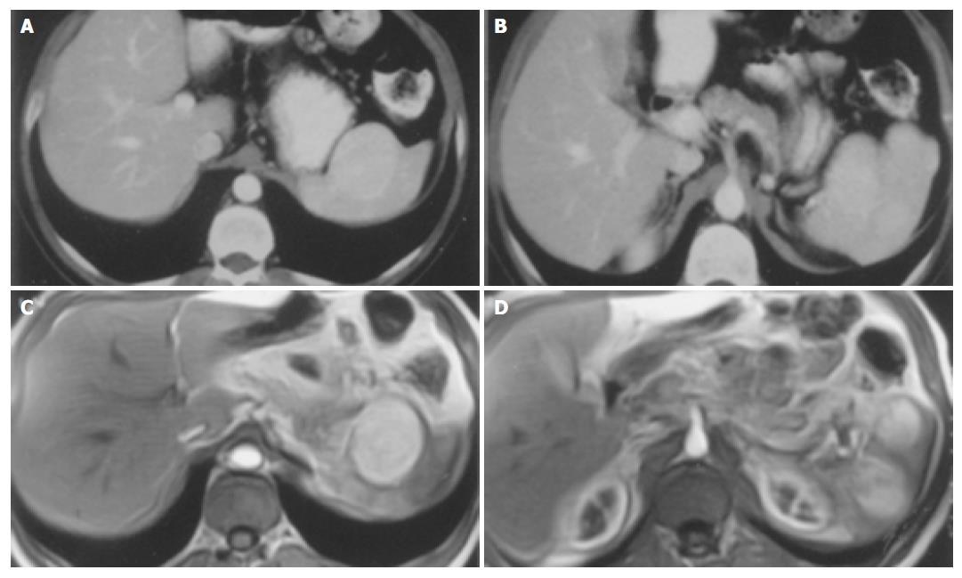
A healthy 31-years-old man presented with a three-year history of early satiety and abdominal discomfort. He had no history of weight loss. Physical and laboratory examinations were normal. Gastroduedonoscopy showed no pathologic finding. Abdominal ultrasonography revealed suspicious nodular lesions in the spleen. Computed tomography and magnetic resonance imaging showed hypervascular and hypodense masses located in the splenic hilum (Figure 1). Maximum size of the masses was 4 cm in diameter.
The patient underwent splenectomy for suspicion of primary or secondary splenic malignancy. During the operation, there were three exophytic nodular lesions in the splenic hilum. In addition, there were also multiple small lesions dark-red in color and 4-5 mm in diameter, resembling to the purpuric lesions macroscopically, on the serosal surface of the splenic flexura of the colon and descending colon. One of them was excised.
Spleen was 10 cm × 8 cm × 7 cm in diameter and weighted 200 g and there were three masses located in the splenic hilum. The gross appearance of the masses showed colour exchange into grayish-brown with a maximum diameter of 3 cm. Tumors were uncapsulated and their borders were irregular. On microscopical examination, there was a tumoral tissue consisting of slightly pleomorphic, atypical cells which were ovoid or spindle in shape. Tumor cells were widespread around the capillary vessels and had fusiform nuclei with prominent nucleolus and eosinophilic cytoplasm (Figures 2A and B). Mitotic activity was not observed. Tumoral cells were stained positively with CD34 and Vimentin, but negatively with lysozyme, cytokeratin, smooth muscle actin, S-100, factor 8-releated antigen, desmin, HMB-45, CD117 and epithelial membrane antigen immunohistochemically. This lesion was reported as hemangiopericytoma.
On histologic examination of the lesion excised from the serosal surface of the splenic colon flexura, there was a tumoral tissue consisting of dilated blood vessels with flattaned endothelium and their lumens were containing erythrocytes (Figure 2C). This lesion was reported as cavernous hemangioma.
Immunohistochemistry of the both specimens (hemang-iopericytoma and cavernous hemangioma) did not display vascular endothelial growth factor (VEGF) expression.
The patient had an uneventfull recovery and discharged on postoperative d 5. He did not take any additional therapy and has been disease free for 36 mo of follow-up.
Despite the advances in imaging techniques, differential diagnosis and management of the splenic hemangiopericytoma may be problematic because of the rarity of the disease. In the present case, ultrasonography, computed tomography, magnetic resonance imaging techniques were inconclusive. In most of the cases, splenectomy seems to be necessary for establishing prompt histologic diagnosis and providing initial treatment of the disease.
Multiple milimetric cavernous hemangiomas of the serosal surfaces of the colon is a rare condition and also splenic hemangiopericytoma is rare. These two distinct tumoral lesions were in the same region, multiple and all three masses in the splenic hilum were growing as exophytic tumors. We hypothesized that there may be triggering substances, which were secreted locally from the tumoral cells, leading paracrine stimulation of the some inducible receptor positive cells in the adjacent tissue. These inducible potential cells had proliferated and developed vascular neoplasia by the effect of these substances. Similarly it has been reported that VEGF may play a role in the pathogenesis of vascular lesions such as cerebral cavernous malformations[10] or cerebral hemangiopericytomas[11]. However VEGF reactivity was negative in both of cavernous hemangioma and splenic hemangiopericytoma in the present case. On the other hand, another substance or mechanism that are not yet clear may play a role in the genesis of this association. We believe that presence of two distinct lesions in the same region was not a co-existence. It may be sequential rather than co-incidental.
Hallen[6] reported that it was difficult to decide whether the splenic hemangiopericytoma was benign or malignant. They also reported that presence of multifocal tumors and mitoses may be an indicator of poor prognosis. In the present case, there were multifocal tumors but mitotic activity was not observed on the histologic examination. Despite multicentric lesions, recurrence has not been described for 36 mo follow-up. Therefore, we think that microscopic features, especially presence of mitosis, may be more important than the presence of multifocal tumors in predicting malign tumor behavior.
In conclusion, splenic hemangiopericytoma and cavernous hemangiomas can be found in the same region and there may be a sequential relationship between these tumoral lesions.
Science Editor Guo SY Language Editor Elsevier HK
| 1. | Stout AP, Murray MR. Hemangiopericytoma: a vascular tumor featuring zimmermann's pericytes. Ann Surg. 1942;116:26-33. [RCA] [PubMed] [DOI] [Full Text] [Cited by in Crossref: 989] [Cited by in RCA: 964] [Article Influence: 53.6] [Reference Citation Analysis (0)] |
| 2. | Enzinger FM, Smith BH. Hemangiopericytoma. An analysis of 106 cases. Hum Pathol. 1976;7:61-82. [RCA] [PubMed] [DOI] [Full Text] [Cited by in Crossref: 625] [Cited by in RCA: 559] [Article Influence: 11.4] [Reference Citation Analysis (0)] |
| 3. | Ciftci AO, Gedikoğlu G, Firat PA, Senocak ME, Büyükpamukçu N. Childhood splenic hemangiopericytoma: a previously unreported entity. J Pediatr Surg. 1999;34:1884-1886. [RCA] [PubMed] [DOI] [Full Text] [Cited by in Crossref: 8] [Cited by in RCA: 8] [Article Influence: 0.3] [Reference Citation Analysis (0)] |
| 4. | Neill JS, Park HK. Hemangiopericytoma of the spleen. Am J Clin Pathol. 1991;95:680-683. [PubMed] |
| 5. | Ferrozzi F, Catanese C, Campani R. Splenic hemangiopericytoma: features with computerized tomography in 2 cases. Radiol Med. 1998;95:122-124. [PubMed] |
| 6. | Hallén M, Parada LA, Gorunova L, Pålsson B, Dictor M, Johansson B. Cytogenetic abnormalities in a hemangiopericytoma of the spleen. Cancer Genet Cytogenet. 2002;136:62-65. [RCA] [PubMed] [DOI] [Full Text] [Cited by in Crossref: 9] [Cited by in RCA: 10] [Article Influence: 0.4] [Reference Citation Analysis (0)] |
| 7. | Guadalajara Jurado J, Turégano Fuentes F, García Menendez C, Larrad Jiménez A, López de la Riva M. Hemangiopericytoma of the spleen. Surgery. 1989;106:575-577. [PubMed] |
| 8. | Hosotani R, Momoi H, Uchida H, Okabe Y, Kudo M, Todo A, Ishikawa T. Multiple hemangiopericytomas of the spleen. Am J Gastroenterol. 1992;87:1863-1865. [PubMed] |
| 9. | Gilsanz C, Garcia-Castaño J, Villalba MV, Vaquerizo MJ, Lopez de la Riva M. [Splenic hemangiopericytoma]. Presse Med. 1995;24:1316. [PubMed] |
| 10. | Jung KH, Chu K, Jeong SW, Park HK, Bae HJ, Yoon BW. Cerebral cavernous malformations with dynamic and progressive course: correlation study with vascular endothelial growth factor. Arch Neurol. 2003;60:1613-1618. [RCA] [PubMed] [DOI] [Full Text] [Cited by in Crossref: 50] [Cited by in RCA: 47] [Article Influence: 2.1] [Reference Citation Analysis (0)] |
| 11. | Hatva E, Böhling T, Jääskeläinen J, Persico MG, Haltia M, Alitalo K. Vascular growth factors and receptors in capillary hemangioblastomas and hemangiopericytomas. Am J Pathol. 1996;148:763-775. [PubMed] |


