Basic Research Open Access
Copyright ©2005 Baishideng Publishing Group Inc. All rights reserved.
World J Gastroenterol. May 28, 2005; 11(20): 3065-3069
Published online May 28, 2005. doi: 10.3748/wjg.v11.i20.3065
Ornithine decarboxylase, mitogen-activated protein kinase and matrix metalloproteinase-2 expressions in human colon tumors
Takahiro Nemoto, Shunichiro Kubota, Department of Physiological Chemistry and Metabolism, Graduate School of Medicine, The University of Tokyo, 7-3-1 Hongo, Bunkyo-ku, Tokyo 113-0033, Japan
Hideyuki Ishida, Nobuo Murata, Daijo Hashimoto, Department of Surgery, Saitama Medical Center, Saitama Medical School, 1981 Tsujido-machi, Kamoda, Kawagoe City, Saitama 350-8550, Japan
Shunichiro Kubota, Department of Life Sciences, Graduate School of Arts and Sciences, The University of Tokyo, 3-8-1 Komaba, Meguro-ku, Tokyo 153-8902, Japan
Author contributions: All authors contributed equally to the work.
Correspondence to: Dr. Shunichiro Kubota, Department of Life Sciences, Graduate School of Arts and Sciences, The University of Tokyo, 3-8-1 Komaba, Meguro-ku, Tokyo 153-8902, Japan. kubota@idaten.c.u-tokyo.ac.jp
Telephone: +81-3-5454-6869 Fax: +81-3-5454-6869
Received: October 8, 2003
Revised: October 9, 2003
Accepted: January 9, 2004
Published online: May 28, 2005

Abstract

AIM: To investigate the expressions of ornithine decarboxylase (ODC), MMP-2, and Erk, and their relationship in human colon tumors.

METHODS: ODC activity, MMP-2 expression, and mitogen-activated protein (MAP) kinase activity (Erk phosphorylation) were determined in 58 surgically removed human colon tumors and their adjacent normal tissues, using [1-14C]-ornithine as a substrate, ELISA assay, and Western blotting, respectively.

RESULTS: ODC activity, MMP-2 expression, and Erk phosphorylation were significantly elevated in colon tumors, compared to those in adjacent normal tissues. A significant correlation was observed between ODC activities and MMP-2 levels.

CONCLUSION: This is the first report showing a significant correlation between ODC activities and MMP-2 levels in human colon tumors. As MMP-2 is involved in cancer invasion and metastasis, and colon cancer overexpresses ODC, suppression of ODC expression may be a rational approach to treat colon cancer which overexpresses ODC.

Key Words: Ornithine decarboxylase, Human colon tumors, mitogen activated protein


INTRODUCTION

Ornithine decarboxylase (ODC) catalyzes the initial step in the biosynthesis of polyamines and ODC activities are associated with malignant status[1]. Overexpression of ODC could lead to cell transformation, tumor invasion, and metastasis[2-4]. A number of studies demonstrated increased expression of ODC in a variety of cancers including colorectal cancer[1].

Invasion and metastasis are a multistep process[5]. Degradation of basement membranes and extracellular matrix is crucial for invasion and metastasis of cancer cells. Basement membranes contain type IV collagen, laminin, heparan sulfate proteoglycan, fibronectin, and other components[5]. Degradation of type IV collagen, a major component of basement membrane, is thought to be a prerequisite for cancer cell invasion[6]. Increased expression or activity of matrix metalloproteinases (MMPs) has been linked to malignancy and tumor cell invasion[7-9]. At least 24 MMPs have been reported so far[7-9]. MMP-2 (72 ku type IV collagenase) which preferentially degrades type IV collagen has been shown to play an important role in invasion and metastasis[5,8]. Several studies have demonstrated increased expression of MMP-2 related to invasion and metastasis of colorectal cancer[10-13].

Mitogen-activated protein (MAP) kinase is a key enzyme of the signal transduction pathways triggered by extracellular signals, including mitogens, growth factors, and cytokines[14-16]. Erk1/2, extracellular signal-regulated kinases that are also termed as p44 and p42 MAP kinases, are ubiquitously expressed. p38 MAP kinase is activated by a variety of cellular stresses, including osmotic shock, inflammatory cytokines, lipopolysaccharides, UV irradiation, and growth factors[17]. Limited information is available concerning MAP kinase expression in human surgically removed colon cancer tissues and its adjacent normal tissues[18]. Hoshino et al[18] showed constitutive activation of MAP kinases in 138 human cancer cell lines, and primary tumor tissues including colon cancer. However, it was reported that the activities of Erk1/2, and p38 MAP kinase were downregulated in the majority of human colon cancers[19]. Sakakura et al[20] also reported infrequent activation of MAP kinase in human colon cancer. Recently, the p38 MAP kinase signaling pathway was shown to be important for the induction of MMP-1 and MMP-9 by extracellular stimuli[21,22]. Therefore, it is imperative to analyze the expression levels of MAP kinases (Erk1/2 and p38) in human surgically removed colon cancer tissues and its adjacent normal tissues.

Colon cancer is one of the most common malignancies in the world and is incurable in its advanced stages[23]. Therefore, it is important to understand the mechanism of invasion and metastasis, and to develop better treatments to prevent or cure metastatic colon cancer. As colon cancer has increased expression levels of ODC, MMP-2, and MAP kinase, these three molecules are the candidate targets for treatment of colon cancer. It is important to clarify a correlation among expressions of these three molecules. We previously found that ODC overexpression in fibroblasts led to cellular transformation and invasion with concomitant induction of MMP-2 and Erk1/2[3]. Little information is available concerning the correlation among ODC, MMP-2, and MAP kinase expressions in cancer in vitro and in vivo. The aim of this study was to investigate the expressions of ODC, MAP kinase, and MMP-2, and further to elucidate the relationship among these expressions in human colon cancer. We discussed the strategies of colon cancer treatment or prevention using α-difluoromethylornithine (DFMO), an irreversible inhibitor of ODC.

MATERIALS AND METHODS
Reagents

Antibodies against phospho-Erk and phospho-p38 MAP kinase were obtained from New England Biolabs (Beverly, MA, USA). MMP-2 ELISA system and DL-[1-14C] ornithine were obtained from Amersham Pharmacia Biotech (Buckinghamshire, UK).

Samples

Paired specimens (tumor and non-cancer tissues adjacent to tumor) were obtained from a total of 58 colon cancer patients who had undergone surgical operation for adenocarcinoma at Saitama Medical University Medical Center. Written informed consent was obtained from all patients before surgical operation. All specimens were immediately stored at -80 °C until use.

ODC enzyme assay

ODC activity was determined using [1-14C]-ornithine as a substrate as described previously[3].

MMP-2 ELISA and Western blot analysis

Tissue lysates were prepared by sonicating tissues in lysis buffer (10 mmol/L Tris-HCl buffer, pH 7.5, containing 1 mmol/L EDTA, 0.5 μg/mL aprotinin, 1 μg/mL leupeptin, and 0.2 mmol/L PMSF) and centrifuged at 12000 g for 10 min at 4 °C. Measurement of protein concentration in supernatant was performed using a Bio-Rad protein assay kit (Hercules, CA, USA). Western blotting was performed as previously described[24]. MMP-2 ELISA was performed according to the manufacturer’s protocol. Quantification of the bands was performed using a NIH image.

Statistical analysis

The results were expressed as mean±SD from three independent experiments. The significance was determined by the paired t-test or Pearson’s test. Statistical analyses were performed using the Statview 4.5 software (Abacus Concepts, Inc., Berkeley, CA, USA).

RESULTS
ODC activity in colon cancer tissues

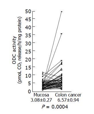
We examined ODC activities in surgically excised human colon cancer (Figure 1). A remarkable increase in ODC activities was seen in colon cancers. Using the paired t-test, a significant increase in ODC activities in colon cancer tissues (6.57±0.94 pmoL CO2 release/h/mg protein) was shown (P = 0.0004 by the paired t-test, n = 58), compared with those observed in adjacent non-cancer tissues (3.08±0.27 pmoL CO2 release/h/mg protein). Increased ODC activity over the normal mean±SD level of colon cancer was seen in 17 of 58 (29.3%) cases.

Figure 1
Figure 1 ODC activity in colon cancer tissues and adjacent normal tissues. ODC activities in colon cancer tissues and adjacent normal tissues were assayed as described in Materials and methods, and the data are shown as mean±SD (pmol CO2 release/h/mg protein).
MMP-2 levels in colon cancer tissues

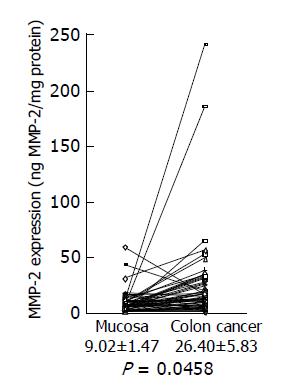
Next, we investigated MMP-2 expression in colon cancer using a MMP-2 ELISA kit. A significant increase in MMP-2 levels was shown in colon cancer tissues (26.40±5.83 ng/mg protein), compared with adjacent non-cancer tissues (9.02±1.47 ng/mg protein) (Figure 2) (P = 0.046 by the paired t-test, n = 58). Increased MMP-2 expression over the level of the normal mean±SD in colon cancer was observed in 28 of 58 (48.3%) cancers.

Figure 2
Figure 2 MMP-2 expression levels in colon cancer tissues and adjacent normal tissues. MMP-2 expression levels in colon cancer tissues and adjacent normal tissues were analyzed by MMP-2 ELISA, and the data are shown as mean±SD (ng MMP-2/mg protein).
Expressions of phosphorylated Erk and p38 MAP kinase in colon cancer tissues

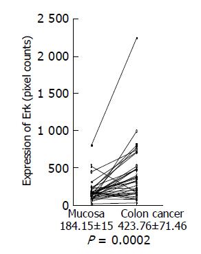
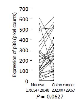
Expression of phosphorylated Erk1/2 and p38 MAP kinase was analyzed by Western blotting using the antibodies against phosphorylated Erk and p38 MAP kinase, respectively. Because of limited amount of tumor tissues, 33 out of 58 samples were used for this experiment. Using the paired t-test, a significant increase in phosphorylated Erk1/2 was shown in colon cancer (423.76±71.46 pixel counts) (P = 0.0002, n = 33), compared with the adjacent tissues (184.15±15.0 pixel counts) (Figure 3). Mean value of phosphorylated p38 MAP kinase had a tendency to increase in colon cancer (232.00±29.67 pixel counts in colon cancer tissues vs 179.54±28.48 pixel counts in adjacent tissues), although P value did not reach a significant level (P = 0.0627) (Figure 4).

Figure 3
Figure 3 Erk expression in colon cancer tissues and adjacent normal tissues. Expression of phosphorylated Erk1/2 in colon cancer tissues and adjacent normal tissues was analyzed by Western blotting using a specific antibody against phosphorylated Erk1/2, quantitated using a NIH image, and shown as mean±SD (pixel counts).
Figure 4
Figure 4 p38 MAP kinase expression in colon cancer tissues and adjacent normal tissues. Expression of phosphorylated p38 MAP kinase in colon cancer tissues and adjacent normal tissues was analyzed by Western blotting using a specific antibody against phosphorylated p38 MAP kinase, quantitated using a NIH image, and are shown as mean±SD (pixel counts).
Correlation between ODC activity, MMP-2 and Erk expressions

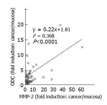
A significant correlation was noted between ODC activity and MMP-2 expression in colon cancer tissues and adjacent normal tissues (r2 = 0.368, P<0.001, n = 58) (Figure 5). Although a correlation between ODC activity and phosphorylated Erk1/2 expression was not found (r2 = 0.028, P = 0.35) in colon cancers (Figure 6), increased expression of phosphorylated Erk1/2 over the level of the normal mean±SD level was observed in 9 of 33 (27.3%) colon cancers (Figure 3). Increased expression of phosphorylated p38 MAP kinase over the level of the normal mean±SD was observed only in 3 of 33 (9.1%) colon cancers (Figure 4).

Figure 5
Figure 5 Relationship between ODC activities and MMP-2 expression in colon cancer tissues. Correlation between ODC activities and MMP-2 expression levels in cancer tissues, compared to those in adjacent normal tissues was shown. Statistical significance was analyzed as described in Materials and methods.
Figure 6
Figure 6 Relationship between ODC activities and Erk expression in colon cancer tissues. Correlation between ODC activities and Erk1/2 expression levels in cancer tissues, compared to those in adjacent normal tissues was shown. Statistical significance was analyzed as described in Materials and methods.
DISCUSSION

In the present study we showed that ODC activities, MMP-2 levels, and Erk1/2 expression had 2.13-, 2.93-, and 2.30-fold increase in colon cancer tissues, respectively, compared to its adjacent normal tissues. Increased ODC activity and MMP-2 levels over the normal mean±SD level in colon cancer were observed in 17 (29.3%) and 28 (48.3%) out of 58 colon cancer patients, respectively. Our results were essentially consistent with the previous report concerning increased ODC activities[1] and increased expression of MMP-2 in colon cancer[10-13]. The data concerning Erk1/2 expression were consistent with the results of a previous report[18], but not with other reports[19,20]. The reason for the inconsistency is not clear. A new finding in the present study was that there was a relationship between expressions of ODC and MMP-2 in colon cancer. We previously demonstrated that ODC overexpression induced MMP-2 and Erk1/2 expressions in fibroblasts[3]. So, it is reasonably assumed that ODC expression induces MMP-2 expression in human colon cancer. A linkage of ODC and MMP-2 expressions very well explains a basis of invasive phenotype of cancers which overexpress ODC.

Metastasis is a multistep process, which includes local invasion, intravasation and extravasation of cancer cells[5]. Major functional contribution of MMPs is facilitation of degradation of basement membranes between primary tumor and distant metastasis sites. As MMP-2 preferentially degrades type IV collagen, a major component of basement membrane, MMP-2 has been thought to play a major role in invasion process[5,8]. Recent evidence indicates that MMPs including MMP-2 play a much broader role in metastasis both before and after the degradation of basement membrane[25]. For example, MMP-2 contributes to the initiation of tumor growth at both primary and metastasis sites. This may involve regulation of growth environment by regulating access to growth factors from the extracellular matrix surrounding the tumor, either directly or via a proteolytic cascade. The role of MMPs in angiogenesis is considered to be very important, as angiogenesis is required for tumor growth and invasion. Recent studies indicated that MMPs, especially MMP-2, played a pivotal role in angiogenesis[26-28]. In fact, mice deficient in MMP-2 exhibited reduced angiogenesis in vivo[29]. As MMP-2 overexpression in colon cancer has been found to be crucial for cancer invasion and metastasis[30], suppression of ODC using DFMO or DFMO in combination with other chemopreventives may be a rational approach to the treatment of colon cancer.

MAP kinase pathways play a major role in converting mitogenic and stress stimuli into nuclear responses. Constitutive activation of the 41-/43-ku MAP kinase (Erk1/2) signaling pathway was shown in human tumors[18]. In the present study we also found increased expression of phosphorylated Erk1/2 in colon cancer. As ODC and Erk1/2 expressions were not correlated in the present study, the data suggested that Erk1/2 expression was not a downstream signal transduction event after ODC expression in human colon cancer. Wang et al[19] reported that the activities of Erk1/2, JNK1 and p38 MAP kinase were downregulated in the majority of human colon cancers. The reason for the discrepancy of our result and their result concerning Erk1/2 is not clear. They discussed that protein kinases other than MAP kinases might play a more crucial role in colon carcinogenesis[19]. In the present study we showed that increased expression of phosphorylated p38 MAP kinase over the level of the normal mean±SD was observed only in 3 of 33 (9.1%) colon cancers. This result might be inconsistent with that of their report[19]. p38 MAP kinase was reported to be involved in other MMP (MMP-1 and MMP-9) expression[21,22]. However, our result and Wang’s result[19], taken together, suggest that p38 MAP kinase may not be involved in carcinogenesis or invasive phenotype or MMP-2 expression in human colon cancer.

As colon cancer is still incurable at advanced stages, new strategies for prevention and treatment are desired[23]. In the 1980s, DFMO was abandoned due to limited efficacy and significant adverse effects such as nausea, vomiting, abdominal pain, diarrhea, and hearing loss in phase II clinical trials[23,31,32]. In the following decade, DFMO was resuscitated as a chemopreventive agent[33,34]. DFMO (0.2-1.0 g/m2/d) effectively inhibited ODC activities in target organs such as the colorectum in patients with a personal or family history of colorectal cancer without side effects such as cytotoxicity[35,36]. DFMO is now being tested in phase II/III trials involving patients at risk for colorectal cancer[23]. Recent studies have shown that DFMO had synergistic reductions in intestinal neoplasia when DFMO was administered with cyclooxygenase inhibitors such as piroxicam or aspirin even as the doses of both agents were reduced as much as 50%[37-41]. Thus, DFMO in combination with other chemopreventive agents should be evaluated for the efficacy against colon cancer. As PD 184352 (MEK inhibitor)[42] or BB-94 (MMP inhibitor)[43] has been available for clinical trial, DFMO in combination of PD 184352 or BB-94 may be a rational approach to the treatment of colon cancers overexpressing ODC.

Footnotes
References
1.  Cohen SS Polyamine metabolism and the promotion of tumor growth. In: Cohen, S.S. (ed.) A guide to the polyamines. New York; Oxford University Press 1998; 296-319.  [PubMed]  [DOI]  [Cited in This Article: ]
2.  Auvinen M, Paasinen A, Andersson LC, Hölttä E. Ornithine decarboxylase activity is critical for cell transformation. Nature. 1992;360:355-358.  [PubMed]  [DOI]  [Cited in This Article: ]  [Cited by in Crossref: 442]  [Cited by in F6Publishing: 463]  [Article Influence: 14.5]  [Reference Citation Analysis (0)]
3.  Kubota S, Kiyosawa H, Nomura Y, Yamada T, Seyama Y. Ornithine decarboxylase overexpression in mouse 10T1/2 fibroblasts: cellular transformation and invasion. J Natl Cancer Inst. 1997;89:567-571.  [PubMed]  [DOI]  [Cited in This Article: ]  [Cited by in Crossref: 57]  [Cited by in F6Publishing: 57]  [Article Influence: 2.1]  [Reference Citation Analysis (0)]
4.  Moshier JA, Dosescu J, Skunca M, Luk GD. Transformation of NIH/3T3 cells by ornithine decarboxylase overexpression. Cancer Res. 1993;53:2618-2622.  [PubMed]  [DOI]  [Cited in This Article: ]
5.  Liotta LA. Tumor invasion and metastases--role of the extracellular matrix: Rhoads Memorial Award lecture. Cancer Res. 1986;46:1-7.  [PubMed]  [DOI]  [Cited in This Article: ]
6.  Chambers AF, Matrisian LM. Changing views of the role of matrix metalloproteinases in metastasis. J Natl Cancer Inst. 1997;89:1260-1270.  [PubMed]  [DOI]  [Cited in This Article: ]  [Cited by in Crossref: 1112]  [Cited by in F6Publishing: 1100]  [Article Influence: 40.7]  [Reference Citation Analysis (0)]
7.  Westermarck J, Kähäri VM. Regulation of matrix metalloproteinase expression in tumor invasion. FASEB J. 1999;13:781-792.  [PubMed]  [DOI]  [Cited in This Article: ]
8.  Yu AE, Murphy AN, Stetler-Stevenson WG.  72 kDa Gelatinase (Gelatinase A): Structure, Activation, Regulation, and Substrate Specificity. In Parks, W.C., and Mecham RP, (ed.) Matrix Metalloproteinases. 1st ed. San Diego: Academic Press 1998; 85-113.  [PubMed]  [DOI]  [Cited in This Article: ]
9.  Nelson AR, Fingleton B, Rothenberg ML, Matrisian LM. Matrix metalloproteinases: biologic activity and clinical implications. J Clin Oncol. 2000;18:1135-1149.  [PubMed]  [DOI]  [Cited in This Article: ]
10.  Levy AT, Cioce V, Sobel ME, Garbisa S, Grigioni WF, Liotta LA, Stetler-Stevenson WG. Increased expression of the Mr 72,000 type IV collagenase in human colonic adenocarcinoma. Cancer Res. 1991;51:439-444.  [PubMed]  [DOI]  [Cited in This Article: ]
11.  Parsons SL, Watson SA, Collins HM, Griffin NR, Clarke PA, Steele RJ. Gelatinase (MMP-2 and -9) expression in gastrointestinal malignancy. Br J Cancer. 1998;78:1495-1502.  [PubMed]  [DOI]  [Cited in This Article: ]  [Cited by in Crossref: 80]  [Cited by in F6Publishing: 91]  [Article Influence: 3.5]  [Reference Citation Analysis (0)]
12.  Liabakk NB, Talbot I, Smith RA, Wilkinson K, Balkwill F. Matrix metalloprotease 2 (MMP-2) and matrix metalloprotease 9 (MMP-9) type IV collagenases in colorectal cancer. Cancer Res. 1996;56:190-196.  [PubMed]  [DOI]  [Cited in This Article: ]
13.  Papadopoulou S, Scorilas A, Arnogianaki N, Papapanayiotou B, Tzimogiani A, Agnantis N, Talieri M. Expression of gelatinase-A (MMP-2) in human colon cancer and normal colon mucosa. Tumour Biol. 2001;22:383-389.  [PubMed]  [DOI]  [Cited in This Article: ]  [Cited by in Crossref: 34]  [Cited by in F6Publishing: 36]  [Article Influence: 1.6]  [Reference Citation Analysis (0)]
14.  Seger R, Krebs EG. The MAPK signaling cascade. FASEB J. 1995;9:726-735.  [PubMed]  [DOI]  [Cited in This Article: ]
15.  Hill CS, Treisman R. Transcriptional regulation by extracellular signals: mechanisms and specificity. Cell. 1995;80:199-211.  [PubMed]  [DOI]  [Cited in This Article: ]  [Cited by in Crossref: 967]  [Cited by in F6Publishing: 955]  [Article Influence: 32.9]  [Reference Citation Analysis (0)]
16.  Mansour SJ, Matten WT, Hermann AS, Candia JM, Rong S, Fukasawa K, Vande Woude GF, Ahn NG. Transformation of mammalian cells by constitutively active MAP kinase kinase. Science. 1994;265:966-970.  [PubMed]  [DOI]  [Cited in This Article: ]  [Cited by in Crossref: 1052]  [Cited by in F6Publishing: 1075]  [Article Influence: 35.8]  [Reference Citation Analysis (0)]
17.  Raingeaud J, Gupta S, Rogers JS, Dickens M, Han J, Ulevitch RJ, Davis RJ. Pro-inflammatory cytokines and environmental stress cause p38 mitogen-activated protein kinase activation by dual phosphorylation on tyrosine and threonine. J Biol Chem. 1995;270:7420-7426.  [PubMed]  [DOI]  [Cited in This Article: ]  [Cited by in Crossref: 1750]  [Cited by in F6Publishing: 1792]  [Article Influence: 61.8]  [Reference Citation Analysis (0)]
18.  Hoshino R, Chatani Y, Yamori T, Tsuruo T, Oka H, Yoshida O, Shimada Y, Ari-i S, Wada H, Fujimoto J. Constitutive activation of the 41-/43-kDa mitogen-activated protein kinase signaling pathway in human tumors. Oncogene. 1999;18:813-822.  [PubMed]  [DOI]  [Cited in This Article: ]  [Cited by in Crossref: 541]  [Cited by in F6Publishing: 521]  [Article Influence: 20.8]  [Reference Citation Analysis (0)]
19.  Wang Q, Ding Q, Dong Z, Ehlers RA, Evers BM. Downregulation of mitogen-activated protein kinases in human colon cancers. Anticancer Res. 2000;20:75-83.  [PubMed]  [DOI]  [Cited in This Article: ]
20.  Sakakura C, Hagiwara A, Shirahama T, Nakanishi M, Yasuoka R, Fujita Y, Inazawa J, Abe T, Kohno M, Yamagishi H. Infrequent activation of mitogen-activated protein kinase in human colon cancers. Hepatogastroenterology. 1999;46:2831-2834.  [PubMed]  [DOI]  [Cited in This Article: ]
21.  Westermarck J, Holmström T, Ahonen M, Eriksson JE, Kähäri VM. Enhancement of fibroblast collagenase-1 (MMP-1) gene expression by tumor promoter okadaic acid is mediated by stress-activated protein kinases Jun N-terminal kinase and p38. Matrix Biol. 1998;17:547-557.  [PubMed]  [DOI]  [Cited in This Article: ]  [Cited by in Crossref: 64]  [Cited by in F6Publishing: 69]  [Article Influence: 2.7]  [Reference Citation Analysis (0)]
22.  Simon C, Goepfert H, Boyd D. Inhibition of the p38 mitogen-activated protein kinase by SB 203580 blocks PMA-induced Mr 92,000 type IV collagenase secretion and in vitro invasion. Cancer Res. 1998;58:1135-1139.  [PubMed]  [DOI]  [Cited in This Article: ]
23.  Umar A, Viner JL, Hawk ET. The future of colon cancer prevention. Ann N Y Acad Sci. 2001;952:88-108.  [PubMed]  [DOI]  [Cited in This Article: ]  [Cited by in Crossref: 25]  [Cited by in F6Publishing: 27]  [Article Influence: 1.2]  [Reference Citation Analysis (0)]
24.  Nemoto T, Kamei S, Seyama Y, Kubota S. p53 independent G(1) arrest induced by DL-alpha-difluoromethylornithine. Biochem Biophys Res Commun. 2001;280:848-854.  [PubMed]  [DOI]  [Cited in This Article: ]  [Cited by in Crossref: 12]  [Cited by in F6Publishing: 13]  [Article Influence: 0.6]  [Reference Citation Analysis (0)]
25.  Chambers AF, Matrisian LM. Changing views of the role of matrix metalloproteinases in metastasis. J Natl Cancer Inst. 1997;89:1260-1270.  [PubMed]  [DOI]  [Cited in This Article: ]
26.  Hiraoka N, Allen E, Apel IJ, Gyetko MR, Weiss SJ. Matrix metalloproteinases regulate neovascularization by acting as pericellular fibrinolysins. Cell. 1998;95:365-377.  [PubMed]  [DOI]  [Cited in This Article: ]  [Cited by in Crossref: 525]  [Cited by in F6Publishing: 545]  [Article Influence: 21.0]  [Reference Citation Analysis (0)]
27.  Stetler-Stevenson WG. Matrix metalloproteinases in angiogenesis: a moving target for therapeutic intervention. J Clin Invest. 1999;103:1237-1241.  [PubMed]  [DOI]  [Cited in This Article: ]  [Cited by in Crossref: 593]  [Cited by in F6Publishing: 590]  [Article Influence: 23.6]  [Reference Citation Analysis (0)]
28.  Werb Z, Vu TH, Rinkenberger JL, Coussens LM. Matrix-degrading proteases and angiogenesis during development and tumor formation. APMIS. 1999;107:11-18.  [PubMed]  [DOI]  [Cited in This Article: ]  [Cited by in Crossref: 116]  [Cited by in F6Publishing: 128]  [Article Influence: 5.1]  [Reference Citation Analysis (0)]
29.  Itoh T, Tanioka M, Yoshida H, Yoshioka T, Nishimoto H, Itohara S. Reduced angiogenesis and tumor progression in gelatinase A-deficient mice. Cancer Res. 1998;58:1048-1051.  [PubMed]  [DOI]  [Cited in This Article: ]
30.  Stetler-Stevenson WG, Liotta LA, Kleiner DE. Extracellular matrix 6: role of matrix metalloproteinases in tumor invasion and metastasis. FASEB J. 1993;7:1434-1441.  [PubMed]  [DOI]  [Cited in This Article: ]
31.  Ajani JA, Ota DM, Grossie VB, Abbruzzese JL, Faintuch JS, Patt YZ, Jackson DE, Levin B, Nishioka K. Evaluation of continuous-infusion alpha-difluoromethylornithine therapy for colorectal carcinoma. Cancer Chemother Pharmacol. 1990;26:223-226.  [PubMed]  [DOI]  [Cited in This Article: ]  [Cited by in Crossref: 10]  [Cited by in F6Publishing: 10]  [Article Influence: 0.3]  [Reference Citation Analysis (0)]
32.  Abeloff MD, Rosen ST, Luk GD, Baylin SB, Zeltzman M, Sjoerdsma A. Phase II trials of alpha-difluoromethylornithine, an inhibitor of polyamine synthesis, in advanced small cell lung cancer and colon cancer. Cancer Treat Rep. 1986;70:843-845.  [PubMed]  [DOI]  [Cited in This Article: ]
33.  Love RR, Carbone PP, Verma AK, Gilmore D, Carey P, Tutsch KD, Pomplun M, Wilding G. Randomized phase I chemoprevention dose-seeking study of alpha-difluoromethylornithine. J Natl Cancer Inst. 1993;85:732-737.  [PubMed]  [DOI]  [Cited in This Article: ]  [Cited by in Crossref: 58]  [Cited by in F6Publishing: 58]  [Article Influence: 1.9]  [Reference Citation Analysis (0)]
34.  Meyskens FL, Emerson SS, Pelot D, Meshkinpour H, Shassetz LR, Einspahr J, Alberts DS, Gerner EW. Dose de-escalation chemoprevention trial of alpha-difluoromethylornithine in patients with colon polyps. J Natl Cancer Inst. 1994;86:1122-1130.  [PubMed]  [DOI]  [Cited in This Article: ]  [Cited by in Crossref: 66]  [Cited by in F6Publishing: 72]  [Article Influence: 2.4]  [Reference Citation Analysis (0)]
35.  Meyskens FL, Gerner EW, Emerson S, Pelot D, Durbin T, Doyle K, Lagerberg W. Effect of alpha-difluoromethylornithine on rectal mucosal levels of polyamines in a randomized, double-blinded trial for colon cancer prevention. J Natl Cancer Inst. 1998;90:1212-1218.  [PubMed]  [DOI]  [Cited in This Article: ]  [Cited by in Crossref: 90]  [Cited by in F6Publishing: 96]  [Article Influence: 3.7]  [Reference Citation Analysis (0)]
36.  Love RR, Jacoby R, Newton MA, Tutsch KD, Simon K, Pomplun M, Verma AK. A randomized, placebo-controlled trial of low-dose alpha-difluoromethylornithine in individuals at risk for colorectal cancer. Cancer Epidemiol Biomarkers Prev. 1998;7:989-992.  [PubMed]  [DOI]  [Cited in This Article: ]
37.  Jacoby RF, Cole CE, Tutsch K, Newton MA, Kelloff G, Hawk ET, Lubet RA. Chemopreventive efficacy of combined piroxicam and difluoromethylornithine treatment of Apc mutant Min mouse adenomas, and selective toxicity against Apc mutant embryos. Cancer Res. 2000;60:1864-1870.  [PubMed]  [DOI]  [Cited in This Article: ]
38.  Nigro ND, Bull AW, Boyd ME. Inhibition of intestinal carcinogenesis in rats: effect of difluoromethylornithine with piroxicam or fish oil. J Natl Cancer Inst. 1986;77:1309-1313.  [PubMed]  [DOI]  [Cited in This Article: ]
39.  Reddy BS, Nayini J, Tokumo K, Rigotty J, Zang E, Kelloff G. Chemoprevention of colon carcinogenesis by concurrent administration of piroxicam, a nonsteroidal antiinflammatory drug with D,L-alpha-difluoromethylornithine, an ornithine decarboxylase inhibitor, in diet. Cancer Res. 1990;50:2562-2568.  [PubMed]  [DOI]  [Cited in This Article: ]
40.  Rao CV, Tokumo K, Rigotty J, Zang E, Kelloff G, Reddy BS. Chemoprevention of colon carcinogenesis by dietary administration of piroxicam, alpha-difluoromethylornithine, 16 alpha-fluoro-5-androsten-17-one, and ellagic acid individually and in combination. Cancer Res. 1991;51:4528-4534.  [PubMed]  [DOI]  [Cited in This Article: ]
41.  Li H, Schut HA, Conran P, Kramer PM, Lubet RA, Steele VE, Hawk EE, Kelloff GJ, Pereira MA. Prevention by aspirin and its combination with alpha-difluoromethylornithine of azoxymethane-induced tumors, aberrant crypt foci and prostaglandin E2 levels in rat colon. Carcinogenesis. 1999;20:425-430.  [PubMed]  [DOI]  [Cited in This Article: ]  [Cited by in Crossref: 66]  [Cited by in F6Publishing: 68]  [Article Influence: 2.7]  [Reference Citation Analysis (0)]
42.  Sebolt-Leopold JS, Dudley DT, Herrera R, Van Becelaere K, Wiland A, Gowan RC, Tecle H, Barrett SD, Bridges A, Przybranowski S. Blockade of the MAP kinase pathway suppresses growth of colon tumors in vivo. Nat Med. 1999;5:810-816.  [PubMed]  [DOI]  [Cited in This Article: ]  [Cited by in Crossref: 729]  [Cited by in F6Publishing: 709]  [Article Influence: 28.4]  [Reference Citation Analysis (0)]
43.  Wojtowicz-Praga S, Low J, Marshall J, Ness E, Dickson R, Barter J, Sale M, McCann P, Moore J, Cole A. Phase I trial of a novel matrix metalloproteinase inhibitor batimastat (BB-94) in patients with advanced cancer. Invest New Drugs. 1996;14:193-202.  [PubMed]  [DOI]  [Cited in This Article: ]  [Cited by in Crossref: 70]  [Cited by in F6Publishing: 77]  [Article Influence: 2.8]  [Reference Citation Analysis (0)]