Copyright
©2005 Baishideng Publishing Group Inc.
World J Gastroenterol. May 28, 2005; 11(20): 3065-3069
Published online May 28, 2005. doi: 10.3748/wjg.v11.i20.3065
Published online May 28, 2005. doi: 10.3748/wjg.v11.i20.3065
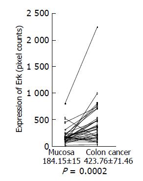
Figure 3 Erk expression in colon cancer tissues and adjacent normal tissues.
Expression of phosphorylated Erk1/2 in colon cancer tissues and adjacent normal tissues was analyzed by Western blotting using a specific antibody against phosphorylated Erk1/2, quantitated using a NIH image, and shown as mean±SD (pixel counts).
- Citation: Nemoto T, Kubota S, Ishida H, Murata N, Hashimoto D. Ornithine decarboxylase, mitogen-activated protein kinase and matrix metalloproteinase-2 expressions in human colon tumors. World J Gastroenterol 2005; 11(20): 3065-3069
- URL: https://www.wjgnet.com/1007-9327/full/v11/i20/3065.htm
- DOI: https://dx.doi.org/10.3748/wjg.v11.i20.3065


