Published online May 21, 2005. doi: 10.3748/wjg.v11.i19.3008
Revised: June 30, 2004
Accepted: August 12, 2004
Published online: May 21, 2005
Various benign and malignant conditions could cause biliary obstruction. Compression of extrahepatic bile duct (EBD) by right hepatic artery was reported as a right hepatic artery syndrome but all cases were compressed EBD from stomach side. Our case compressed from dorsum was not yet reported, so it was thought to be a very rare case. We present here the first case of bile duct obstruction due to the compression of EBD from dorsum by right hepatic artery.
- Citation: Miyashita K, Shiraki K, Ito T, Taoka H, Nakano T. The right hepatic artery syndrome. World J Gastroenterol 2005; 11(19): 3008-3009
- URL: https://www.wjgnet.com/1007-9327/full/v11/i19/3008.htm
- DOI: https://dx.doi.org/10.3748/wjg.v11.i19.3008
In the biliary legion, anatomic variation is relatively common. Arterial anomalies are not infrequent findings during biliary surgery and variations in the positions of hepatic arteries[1-5] are well known. However, it has been rarely reported that the extrahepatic bile duct (EBD) is compressed by the vessels of hepatobiliary lesion.
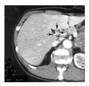
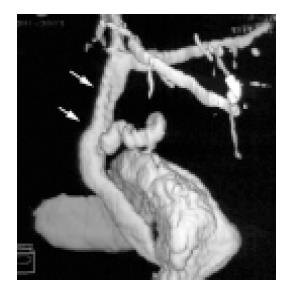
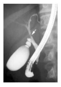
A 55-year-old Japanese man with an unremarkable medical and family history was admitted for fever and body weight loss. On laboratory examination, alkaline phosphatase was 1403 IU/L (normal range: 120-340 IU/L) and gamma-glutamyl transpeptidase was 130 IU/L (normal range: 8-60 IU/L). An abdominal computed tomography (CT) showed compression from the dorsum and stenosis of the EBD (Figure 1: thick arrow head, EBD; Figure 3) by the right hepatic artery (Figure 1: thin arrow head, anterior branch and posterior branch), but did not reveal a mass or lymph node swelling around the EBD. Endoscopic retrograde cholangiography (ERC) showed a stenotic lesion of the common hepatic duct (Figure 2). An intraductal ultrasonography and biopsy were performed transpapillary, but no malignant finding was reported. Drainage of the bile duct using an endoscopic nasobiliary drainage tube improved fever and normalized enzymes. The resultant diagnosis was cholangitis due to compression of the EBD by the right hepatic artery from the posterior side. As a treatment, the stenotic site of the common hepatic duct was removed and end-to-side hepaticojejunostomy was performed. The operation showed that the right hepatic artery had crossed EBD from posterior side of EBD and compressed it. Pathologic examination showed no malignancy or inflammatory cell invasion, but demonstrated sclerosis and severe tortuosity of right hepatic artery. The post-operative course was uneventful and the patient has been well for 1 year after the operation.
Compression of the EBD by the right hepatic artery has been reported as a right hepatic artery syndrome, but in all six cases reported, the EBD was compressed from the stomach side. In particular in this patient, the right hepatic artery ran across the back of the bile duct, which is a standard variation. It is possible that the etiology of the obstructive jaundice in this patient was that the common hepatic artery was short and proximal ramification of the right hepatic artery of the anterior segment branch and posterior segment branch or severe tortuosity of the right hepatic artery by arterial sclerosis.
Science Editor Guo SY Language Editor Elsevier HK
| 1. | Chung JP, Kim KW, Chi HS, Lee SI, Shin ET, Cho JH, Lee HW, Kang JK, Park IS. Obstructive jaundice due to compression of the common hepatic duct by right hepatic artery--a case associated with the absence of the lateral segment of the left hepatic lobe. Yonsei Med J. 1994;35:231-238. [RCA] [PubMed] [DOI] [Full Text] [Cited by in Crossref: 13] [Cited by in RCA: 12] [Article Influence: 0.4] [Reference Citation Analysis (0)] |
| 2. | Tsuchiya R, Eto T, Harada N, Yamamoto K, Matsumoto T, Tsunoda T, Yamaguchi T, Noda T, Izawa K. Compression of the common hepatic duct by the right hepatic artery in intrahepatic gallstones. World J Surg. 1984;8:321-326. [RCA] [PubMed] [DOI] [Full Text] [Cited by in Crossref: 17] [Cited by in RCA: 18] [Article Influence: 0.4] [Reference Citation Analysis (0)] |
| 3. | Kumada S, Nakano H, Kitamura K, Watahiki H, Takeda I, Ozawa H, Hachisuga K. A case of the obstructive jaundice due to compression of the common hepatic duct by right hepatic artery (right hepatic artery syndrome) (in Japanese). Biliary Tract Pancreas. 1981;2:533. |
| 4. | Luttwak EM, Schwartz A. Jaundice due to obstruction of the common duct by aberrant artery: demonstration of celiac anomaly by translumbar aortography and simultaneous choledochogram. Ann Surg. 1961;153:134-137. [RCA] [PubMed] [DOI] [Full Text] [Cited by in Crossref: 14] [Cited by in RCA: 15] [Article Influence: 0.6] [Reference Citation Analysis (0)] |
| 5. | Hunt AH. Compression of the common bile duct by an enlarging collateral vein in a case of portal hypertension. Br J Surg. 1965;52:636-637. [RCA] [PubMed] [DOI] [Full Text] [Cited by in Crossref: 29] [Cited by in RCA: 23] [Article Influence: 0.8] [Reference Citation Analysis (0)] |


