Published online Oct 1, 1995. doi: 10.3748/wjg.v1.i1.37
Revised: August 5, 1995
Accepted: September 9, 1995
Published online: October 1, 1995
AIM: To investigate the pattern of distribution and ontogeny of motilin (MTL) in the human fetus.
METHODS: MTL concentrations were determined systematically, using radioimmunoassay, in the tissues of the central nervous system (CNS, six regions) and the digestive system (20 regions) in human fetuses.
RESULTS: The results showed a wide distribution of MTL in the tissues but at different concentrations. The MTL concentrations in the digestive system tended to be higher than those in the CNS, and the concentrations increased with the fetal age, especially in the digestive system. High concentrations of MTL were found in hypophysis (the earliest MTL generation region in the CNS), spinal cord and cerebellum; however, there were no significant differences among them. In the digestive system, MTL was detectable as early as 16th week in jejunum, duodenum, ileum, stomach, transverse colon, sigmoid colon, and rectum, while in other regions MTL was not detec until after the 23rd week. By the 24th week, MTL showed an adult distribution pattern in all of the tissues. In the digestive system tissue of different fetal ages, the highest MTL concentration was found in jejunum, followed by duodenum and ileum, and MTL was positively correlated with the fetal age. The MTL concentration in ileum was about 6-7 times higher than in the CNS. In addition, we detected MTL at a considerable concentration in the appendix for the first time.
CONCLUSION: The findings might help reveal distribution and ontogeny of MTL in fetuses.
- Citation: Huang YX, Xu CF, Liao H, Wang QL. Genesis and distribution of motilin in the human fetus. World J Gastroenterol 1995; 1(1): 37-40
- URL: https://www.wjgnet.com/1007-9327/full/v1/i1/37.htm
- DOI: https://dx.doi.org/10.3748/wjg.v1.i1.37
Motilin (MTL) is a gastrointestinal hormone which stimulates gastrointestinal tract motility. Despite rapid progress in the MTL research, especially regarding molecular biology and receptor theory, the studies of embryogenesis of MTL have been limited to intestinal organs such as duodenum, jejunum, ileum, and colon. Furthermore, there has been no report on the MTL genesis in the central nervous system (CNS) in human fetus[1]. In our present study, MTL concentrations were determined in the tissues of both CNS and digestive system of human fetuses age 16 wk to 40 wk.
Twelve normal fetuses (5 males and 7 females) in water bag were obtained from healthy pregnant women by labor induction and were stored in a freezer (-32 °C) for no more than one month. Full ethical permission was obtained for this study. Deduced from the time of the last menstrual period of the pregnant women, the fetuses were divided into three groups, 16–23 wk, 24–31 wk, and 32–40 wk.
Fetuses were defrosted, and the esophagus, stomach, duodenum, jejunum, ileum, colon, appendix and gall bladder were dissected under direct vision at 10 °C. The organ lengths were measured, and the tissue specimens were taken from each organ. The blood was removed, and the specimen put in vials which were immediately placed on crushed ice. The samplings from the medullary bulb, cerebral cortex, hypothalamus, cerebellum, hypophysis and spinal cord were taken out as previously described. After all the tissue specimens (20 regions in the gastrointestinal tract and six regions in the CNS) had been taken, they were stored in a freezer (-32 °C).
Tissues weighing 20–100 mg were taken from the cerebrospinal cord and each organ in the gastrointestinal tract. The tissues were defrosted, and 1 mL of 2 mol/L iced acetic acid was added. After been plunged into vigorously boiling water for 10 min, the tissues were cooled down to 4 °C and then homogenized for 10 min. After centrifugation at 3500 rpm for 20 min at 4 °C, the supernatant was collected. Just before the radioimmunoassay (RIA) assay, the supernatant was centrifuged for another 10 min for further purification.
MTL was determined using a non-equilibrium RIA with motilin antibody. The kit was provided by the General Hospital of the People’s Liberation Army. The specimens were assayed in duplicate.
The data were analyzed using the statistical analysis of variance and correlation.
The MTL concentrations in cerebrospinal tissue of different fetal ages are given in Table 1. Hypophysis was the first region to secrete MTL as early as the 16th week while in other regions MTL was not detectable until after a 23rd week. MTL concentrations in human fetal cerebrospinal tissues, hypophysis, cerebellum, and spinal cord were high. However, there was no significant difference in the MTL concentration between the cerebrospinal tissues assayed (P > 0.05).
| Region | Fetal age (wk) | ||
| 16-23 | 24-31 | 32-40 | |
| Cerebral cortex | UD | 0.222 ± 0.052 | 0.967 ± 0.738 |
| Hypothalamus | UD | 0.347 ± 0.152 | 1.015 ± 0.634 |
| Hypophysis | 0.884 ± 0.417 | 0.599 ± 0.408 | 1.678 ± 1.404 |
| Cerebellum | UD | 0.276 ± 0.116 | 1.272 ± 1.101 |
| Medullary bulb | UD | 0.357 ± 0.068 | 1.086 ± 0.749 |
| Spinal cord | UD | 0.270 ± 1.131 | 1.611 ± 1.019 |
The MTL concentrations in digestive tissues of different fetal ages are shown in Table 2. In the digestive system, MTL was detectable before the 16th week in jejunum, duodenum, ileum, stomach, colon, and rectum, whereas in other regions it was detected no earlier than the 23rd week. It was not until the 24th week that the fetuses displayed a similar distribution pattern of MTL to that of adults. MTL levels in both the digestive system and the CNS increased with the age of the fetuses, and the MTL level in the former tended to be elevated more significantly than that of the latter.
| Region | Fetal age (week) | ||
| 16-23 | 24-31 | 32-40 | |
| Esophagus | UD | 0.801 ± 0.483 | 1.441 ± 0.896 |
| Cardia | UD | 0.404 ± 0.239 | 1.775 ± 0.968 |
| Stomach | 0.125 ± 0.071 | 0.618 ± 0.537 | 1.966 ± 1.441 |
| Pylorus | UD | 0.745 ± 0.724 | 1.591 ± 0.753 |
| Duodenum | 1.562 ± 0.009 | 3.764 ± 2.471 | 4.656 ± 2.480 |
| Jejunum | 2.302 ± 0.127 | 3.947 ± 2.729 | 8.488 ± 4.927 |
| Ileum | 0.880 ± 0.173 | 1.066 ± 0.641 | 2.420 ± 1.769 |
| Cecum | UD | 1.119 ± 0.379 | 1.726 ± 0.636 |
| Appendix | UD | 0.688 ± 0.096 | 1.632 ± 0.787 |
| Colon | 0.250 ± 0.083 | 1.054 ± 0.363 | 2.286 ± 1.745 |
| Rectum | 0.112 ± 0.037 | 0.719 ± 0.461 | 2.318 ± 1.033 |
| Gallbladder | UD | 0.457 ± 0.077 | 1.164 ± 0.397 |
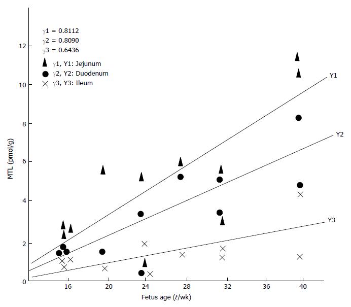
In all the digestive system tissues of different fetal ages, the highest MTL concentration was found in jejunum, followed by duodenum, ileum, and colon. The concentrations in the descending part of the duodenum, jejunum, and colon were positively correlated with the age of the fetuses (P < 0.01 and P < 0.05, respectively) (Figure 1). In addition, for the first time, MTL was found in the appendix.
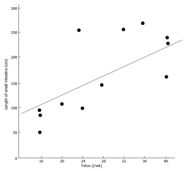
The length of the small intestine (including jejunum and ileum) was closely correlated with the age of the fetuses (r = 0.0056, P < 0.01, Figure 2).
In the cerebrospinal tissues, hypophysis was the first to secrete MTL (16th week), whereas in other regions it was not until 24th week. The MTL secreting cells in the adenohypophysis might be responsible for the early secretion of MTL in hypophysis[2]. The later secretion of MTL in cerebellum and other central nervous tissue suggests that the cells in adenohypophysis might secrete MTL earlier than the Purkinje cells.
MTL in fetuses could be seen as a type of a cerebral enteropeptide, for it is widely distributed in the CNS tissue, especially in hypophysis, cerebellum, and spinal cord, and it exists at various levels in the hypothalamus, medullary bulb, and the cerebral cortex. Although there was no significant difference between these regions, the MTL concentration in hypophysis was twice that of cerebral cortex which might be related to the density distribution of the MTL secreting cells. However, further confirmation is necessary. The MTL concentration in the CNS increased with the fetal age and reached the peak level by the 40th week. This might be due to the gradual maturation of the fetal CNS as the fetus developed, the MTL secreting cells in the CNS increase in numbers and improve in function.
MTL in CNS was different from cholecystokinin and vasoactive intestinal peptide in the subcellular distribution. Rather than located in a vesicle, MTL was located in the nucleus[3]. In the early CNS developmental period, MTL might act either as a chemical signal or as a nutrient for cell migration and linkage between specific neurons and their target cells.
The regions in which MTL was detectable before the 16th week were jejunum, duodenum, ileum, stomach, transverse colon, rectum and sigmoid colon while in other regions it was not detectable until after the 16th week. However, there was an adult distribution pattern in all regions by the 24th week which is in agreement with the 20-24 wk adult distribution pattern of most other gastrointestinal hormones[4]. The genesis of MTL in the majority of gastrointestinal system regions was earlier than that in the CNS.
MTL was widely distributed in the gastrointestinal system. In our present study, it was detected in all of the 20 regions, though at different levels. In all age groups, the highest MTL concentration was noted in jejunum tissues which was twice that in the duodenum, three times that in ileum and colon, six times that in the esophagus and eight times that of a gall bladder. The result confirmed the immunohistochemical report that the most densely distributed region of the motilin-immunopositive cells was the upper segment of the small intestine. MTL concentration in the gastrointestinal system had a tendency to increase with the fetal age and reached the peak level by the 40th week. Having detected MTL of considerable concentration in the appendix for the first time, we deduced that motilin-immunopositive cells might exist in the mucosa of the appendix where appendix potentially acts as a secretory organ. MTL was present in the gall bladder tissues, but was hardly detectable in bile, suggesting a lack of secretion of MTL by the gall bladder tissue into bile, and if any, it was decomposed by an enzyme in bile soon after MTL was secreted. MTL concentrations in the fetal duodenum, jejunum, and ileum were positively correlated with the fetal age. Thus, the assay of MTL concentrations in these regions was of great value in assessing the development of fetal small intestine.
In short, MTL has a wide distribution in both the gastrointestinal and cerebrospinal tissue, with the MTL concentrations in the former were higher than those in the latter. The most remarkable discovery was the MTL concentration in jejunum was about 6-7 times that in the CNS[5].
Presented at the Chinese/Japanese Medical Conference, Beijing, China, l November 1992.
Original title: China National Journal of New Gastroenterology (1995-1997) renamed World Journal of Gastroenterology (1998-).
S- Editor: Filipodia L- Editor: Jennifer E- Editor: Zhang FF
| 1. | Bryant MG, Buchan AM, Gregor M, Ghatei MA, Polak JM, Bloom SR. Development of intestinal regulatory peptides in the human fetus. Gastroenterology. 1982;83:47-54. [PubMed] |
| 2. | Loftus CM, Nilaver G, Beinfeld MC, Post KD. Motilin-related immunoreactivity in mammalian adenohypophysis. Neurosurgery. 1986;19:201-204. [PubMed] |
| 3. | Korchak DM, Nilaver G, Beinfeld MC. The development of motilin-like immunoreactivity in the rat cerebellum and pituitary as determined by radioimmunoassay. Neurosci Lett. 1984;48:267-272. [RCA] [PubMed] [DOI] [Full Text] [Cited by in Crossref: 14] [Cited by in RCA: 15] [Article Influence: 0.4] [Reference Citation Analysis (0)] |
| 4. | Bloom SR, Polak JM. Gut hormone overview. Gut hormones. Edinburgh: Chruchill livingstone 1978; 3-18. |
| 5. | Poitras P, Trudel L, Lahaie RG, Pomier-Layrargue G. Motilin-like-immunoreactivity in intestine and brain of dog. Life Sci. 1987;40:1391-1395. [RCA] [PubMed] [DOI] [Full Text] [Cited by in Crossref: 21] [Cited by in RCA: 19] [Article Influence: 0.5] [Reference Citation Analysis (0)] |


