Copyright
©The Author(s) 2003.
World J Gastroenterol. Aug 15, 2003; 9(8): 1840-1843
Published online Aug 15, 2003. doi: 10.3748/wjg.v9.i8.1840
Published online Aug 15, 2003. doi: 10.3748/wjg.v9.i8.1840
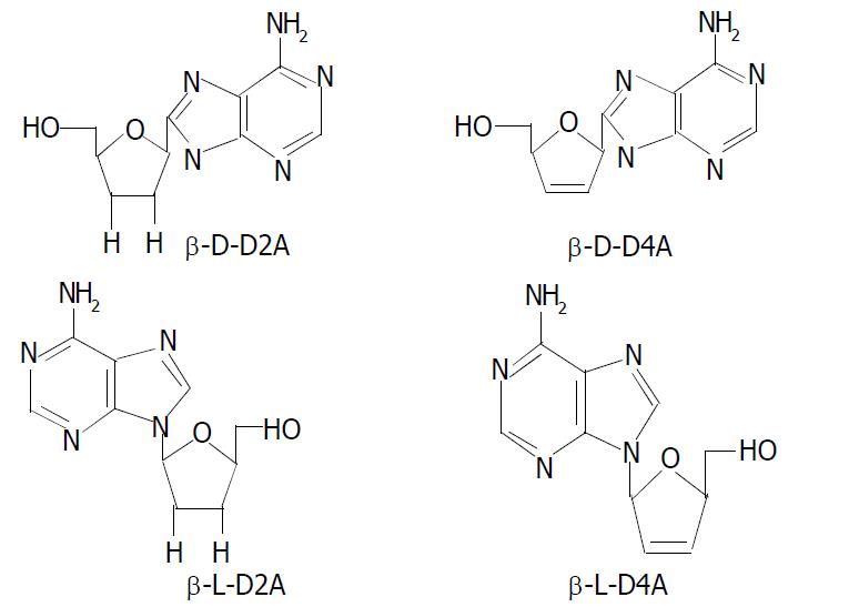
Figure 1 Structures of β-L-D4A and analogues.
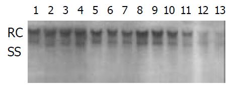
Figure 2 Inhibition of isomers of D4A and D2A on HBV DNA replication in supernatant.
Lane 1 as negative control; lanes 2, 3, 4 as D-D2A at 2 μM, 4 μM and 8 μM, respectively; lanes 5, 6, 7 as L-D2A at 2 μM, 4 μM and 8 μM, respectively; lanes 8, 9, 10 as D-D4A at 2 μM, 4 μM and 8 μM, respectively; lanes 11, 12, 13 as L-D4A at 2 μM, 4 μM and 8 μM, respectively. RC: relaxed circular HBV DNA; SS: single-stranded HBV DNA.
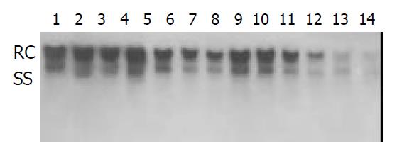
Figure 3 Inhibition on replication of extracellular HBV DNA by β-L-D4A.
lanes 1, 2 as negative control; lanes 3, 4 as lamivudine at 1 μM; lanes 5, 6 as β-L-D4A at 10 μM; lanes 7, 8 as β-L-D4A at 2 μM; lanes 9, 10 as β-L-D4A at 0.4 μM; lanes 11, 12 as β-L-D4A at 0.08 μM; lanes 13, 14 as blank control (HepaG2). RC: relaxed circular HBV DNA; SS: single-stranded HBV DNA.
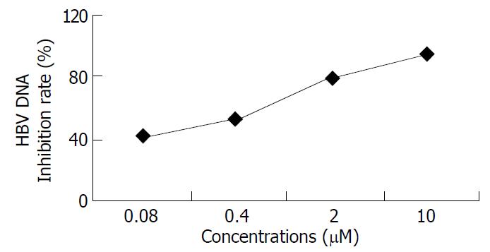
Figure 4 Inhibition on replication of extracellular HBV DNA by β-L-D4A.
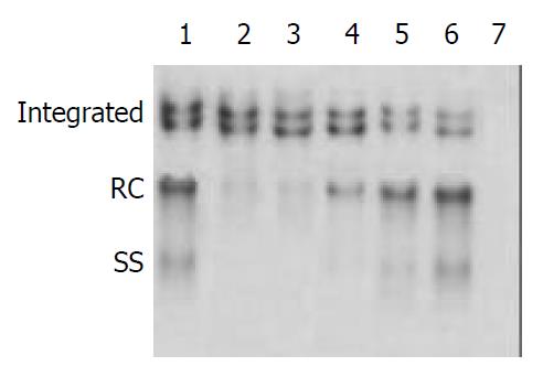
Figure 5 Inhibition on replication of intracellular HBV DNA by β-L-D4A.
lane 1 as negative control; lane 2 as lamivudine at 1 μM; lane 3 as β-L-D4A at 10 μM; lane 4 as β-L-D4A at 2 μM; lane 5 as β-L-D4A at 0.4 μM; lane 6 as β-L-D4A at 0.08; lane 7 as blank control (HepaG2). RC: relaxed circular HBV DNA; SS: single-stranded HBV DNA.
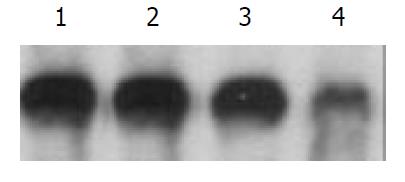
Figure 6 Toxicity of mitochondrial DNA.
lane 1 as blank control; lane 2 as β-L-D4A at 0.4 μM; lane 3 as β-L-D4A at 10 μM; lane 4 as ddC at 0.4 μM.
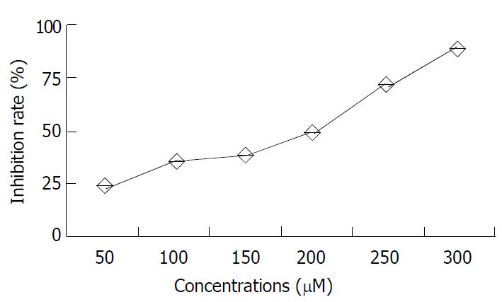
Figure 7 Effect of β-L-D4A on cell growth curve.
- Citation: Wu JM, Lin JS, Xie N, Liang KH. Inhibition of hepatitis B virus by a novel L-nucleoside, β-L-D4A and related analogues. World J Gastroenterol 2003; 9(8): 1840-1843
- URL: https://www.wjgnet.com/1007-9327/full/v9/i8/1840.htm
- DOI: https://dx.doi.org/10.3748/wjg.v9.i8.1840


