Copyright
©The Author(s) 2003.
World J Gastroenterol. Oct 15, 2003; 9(10): 2284-2288
Published online Oct 15, 2003. doi: 10.3748/wjg.v9.i10.2284
Published online Oct 15, 2003. doi: 10.3748/wjg.v9.i10.2284
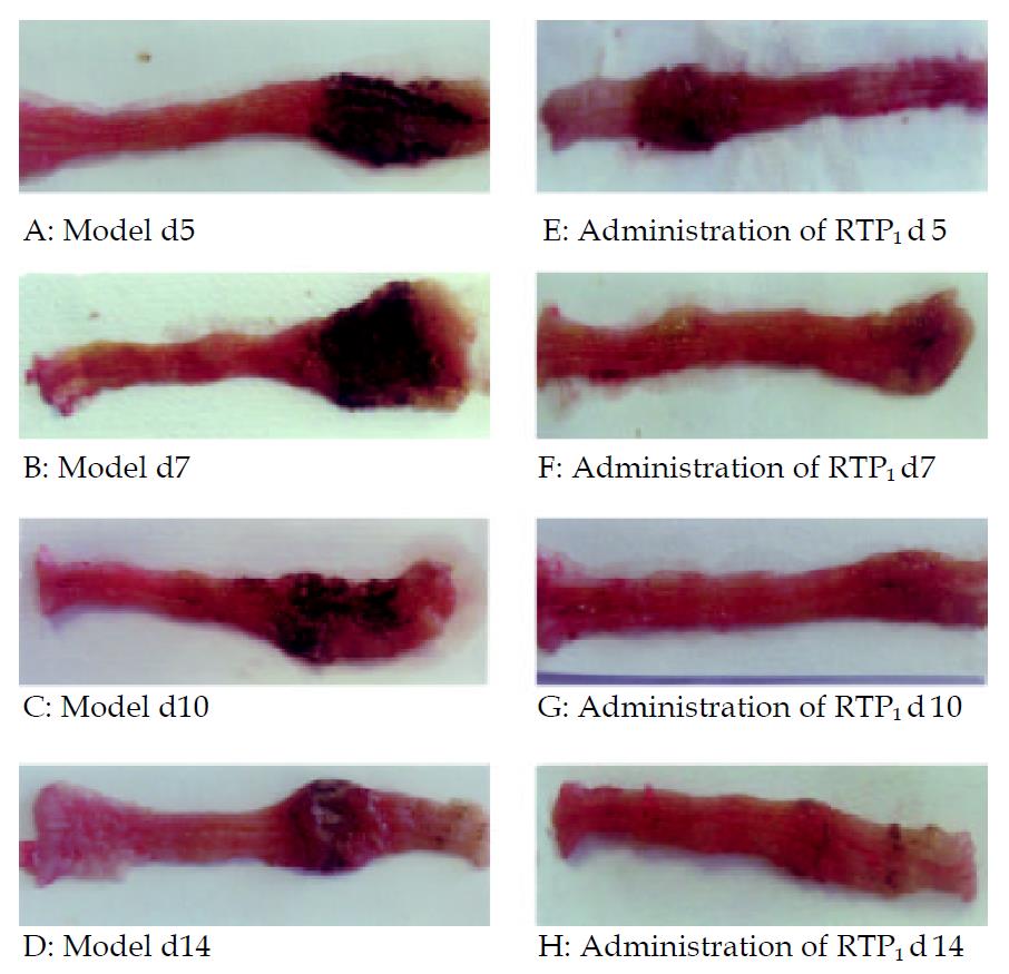
Figure 1 Photographs of rats colon.
A-D. TNBS-induced colitis with gross enlargement of the colon and large ulcers. E-H. TNBS with RTP1, smaller ulcer area and reduced colon mass.
Figure 2 Western-blot analysis of CD4+ T cells isolated from me-senteric lymphoid node.
Left: Saline, Middle: RTP1, Right: TNBS.
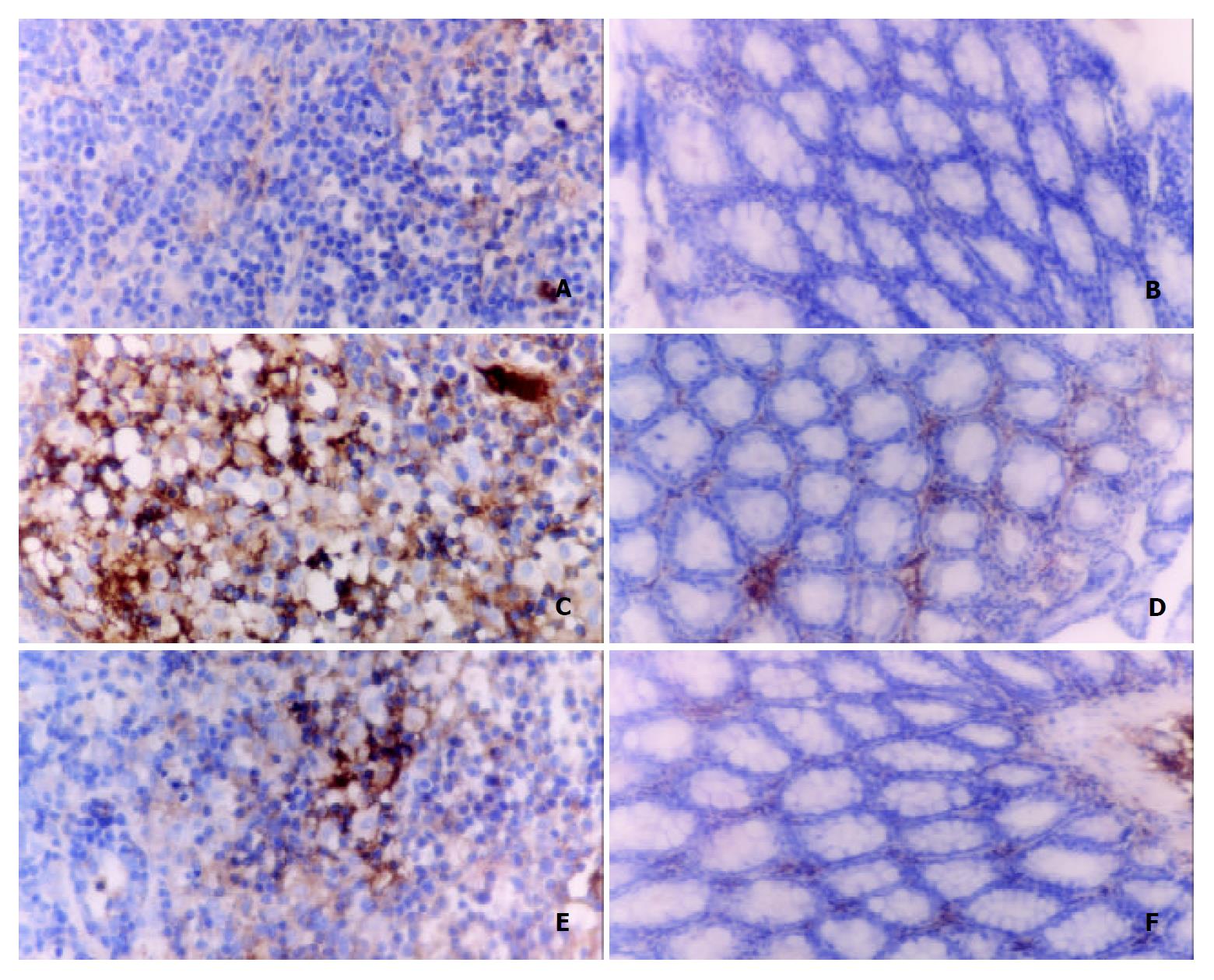
Figure 3 Immunohistochemical analysis on CD4+ lymphocytes in mesenteric lymphoid nodes and colon tissues SABC × 400.
A, C, E: Mesenteric lymphoid nodes. B, D, F: Colon tissues.
-
Citation: Liu L, Wang ZP, Xu CT, Pan BR, Mei QB, Long Y, Liu JY, Zhou SY. Effects of
Rheum tanguticum polysaccharide on TNBS -induced colitis and CD4+ T cells in rats. World J Gastroenterol 2003; 9(10): 2284-2288 - URL: https://www.wjgnet.com/1007-9327/full/v9/i10/2284.htm
- DOI: https://dx.doi.org/10.3748/wjg.v9.i10.2284


