Copyright
©The Author(s) 2003.
World J Gastroenterol. Oct 15, 2003; 9(10): 2261-2265
Published online Oct 15, 2003. doi: 10.3748/wjg.v9.i10.2261
Published online Oct 15, 2003. doi: 10.3748/wjg.v9.i10.2261
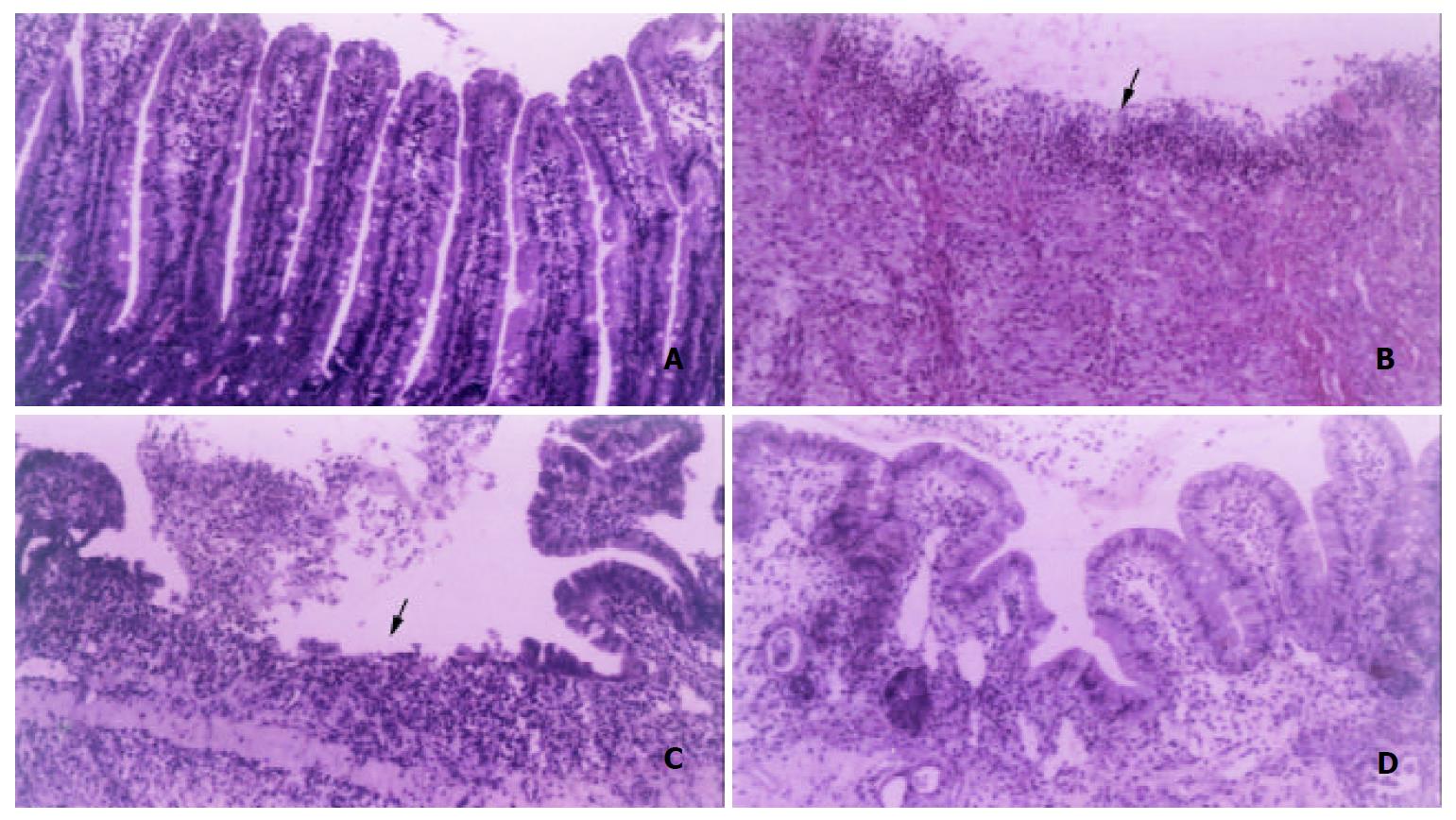
Figure 1 Representative micrographs of duodenal mucosa stained by haematoxylin and eosin were selected from 4 sectioned samples per group.
A: control rats on day 0 (× 200), B: duodenal ulcer rats without EGF on day 1 (× 100), C: duodenal ulcer rats without EGF on day 5 (× 100), D: duodenal ulcer rats with EGF on day 5 (× 200). Arrow represents the discontinuous lining of the duodenal mucosa.
- Citation: Chao JC, Liu KY, Chen SH, Fang CL, Tsao CW. Effect of oral epidermal growth factor on mucosal healing in rats with duodenal ulcer. World J Gastroenterol 2003; 9(10): 2261-2265
- URL: https://www.wjgnet.com/1007-9327/full/v9/i10/2261.htm
- DOI: https://dx.doi.org/10.3748/wjg.v9.i10.2261


