Copyright
©The Author(s) 2002.
World J Gastroenterol. Dec 15, 2002; 8(6): 994-998
Published online Dec 15, 2002. doi: 10.3748/wjg.v8.i6.994
Published online Dec 15, 2002. doi: 10.3748/wjg.v8.i6.994
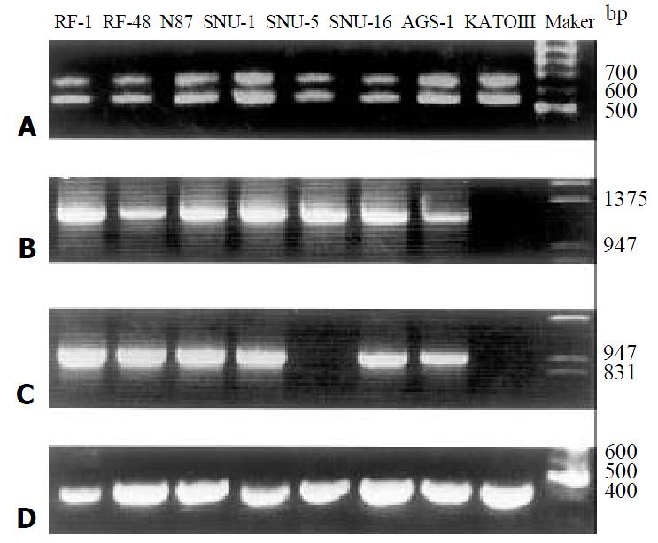
Figure 1 Detection of expression of VEGF and VEGFR in eight gastric carcinoma cell lines by RT-PCR.
A. VEGF was ampli-fied using primers designed to detect all known splicing vari-ants and two isoforms 531 bp and 663 bp corresponding to VEGF121 and VEGF165 were obtained in all cell lines. B. Flt-1 of the expected size (1212 bp) was amplified in all cell lines except KATOIII. C. KDR of the expected size (927 bp) was amplified in all cell lines except SNU-5 and KATOIII. D. GAPDH was amplified in each cell line as a positive control for RT-PCR.
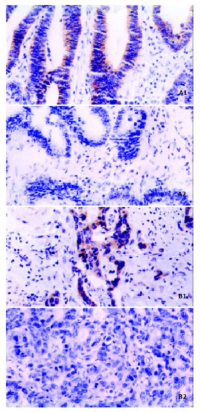
Figure 2 Immunohistochemical analysis of Flt-1 and KDR on gastric carcinoma specimens ( × 400).
A. Expression of Flt-1. B. Expression of KDR. below sections are respectively nega-tive controls of the left-hand side sections of the same specimens.
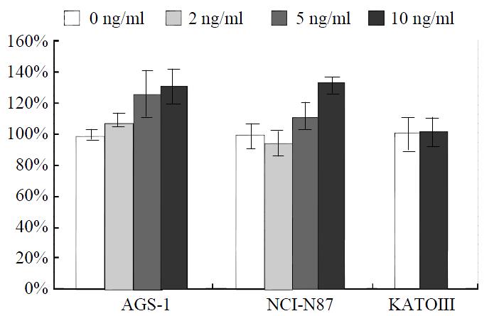
Figure 3 Effects of recombinant human VEGF165 on prolifera-tion of AGS-1, NCI-N87 and KATOIIIcells.
The cells were treated with concentrations of VEGF165 indicated, and their vi-ability was assessed using MTT and expressed as mean per-centage of the untreated controls ± SE (n = 4).
- Citation: Zhang H, Wu J, Meng L, Shou CC. Expression of vascular endothelial growth factor and its receptors KDR and Flt-1 in gastric cancer cells. World J Gastroenterol 2002; 8(6): 994-998
- URL: https://www.wjgnet.com/1007-9327/full/v8/i6/994.htm
- DOI: https://dx.doi.org/10.3748/wjg.v8.i6.994


