Copyright
©The Author(s) 2002.
World J Gastroenterol. Jun 15, 2002; 8(3): 515-519
Published online Jun 15, 2002. doi: 10.3748/wjg.v8.i3.515
Published online Jun 15, 2002. doi: 10.3748/wjg.v8.i3.515
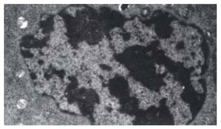
Figure 1 The apoptotic cells in 40 μg·mL-1 IH764-3 for 48 hr (TEM 10000 ×) Apoptotic cells became small, the chromatins condensed
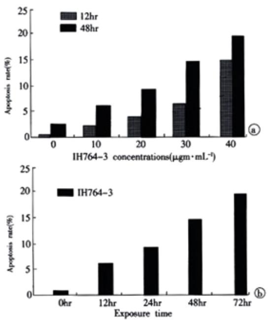
Figure 2 apoptosis rates of HSCs exposed to IH764-3.
(A) dose-dependent; (B) time-dependent with the concentration of 30 μg·mL-1 of IH764-3.
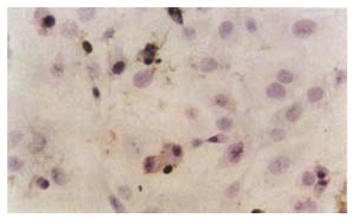
Figure 3 The apoptotic cells in 30 μg·mL-1 IH764-3 for 48 hr (TUNEL10 × 40)
-
Citation: Zhang XL, Liu L, Jiang HQ. Salvia miltiorrhiza monomer IH764-3 induces hepatic stellate cell apoptosis
via caspase-3 activation. World J Gastroenterol 2002; 8(3): 515-519 - URL: https://www.wjgnet.com/1007-9327/full/v8/i3/515.htm
- DOI: https://dx.doi.org/10.3748/wjg.v8.i3.515


