Copyright
©The Author(s) 2002.
World J Gastroenterol. Apr 15, 2002; 8(2): 253-257
Published online Apr 15, 2002. doi: 10.3748/wjg.v8.i2.253
Published online Apr 15, 2002. doi: 10.3748/wjg.v8.i2.253
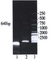
Figure 1 Identification of recombinant plasmids digested with restriction enzymes (Hind III and Cla I) 1: pLncx plasmid digested with Hind III and Cla I; 2: pLncx-Endo plasmid digested with Hind III and Cla I; 3: DNA Marker.
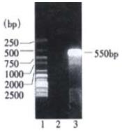
Figure 2 Analysis of PCR product of SMMC7721 transferred with pLncx-endo by 1% agarose gel electrophoresis.
1: DNA Marker; 2: PCR product of SMMC7721 cell DNA transferred with pLncx; 3: PCR product of SMMC7721 cell DNA transferred with pLncx-endo
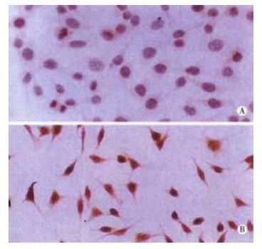
Figure 3 Expression of human endostatin-HA fusion protein in endostatin-transfected cells.
Anti-HA monocolonal antibody was applied to SMMC7721 transferred with pLncx; A: and SMMC7721 transferred with pLncx-endo; B: followed by a HRP-conjugated secondary antibody. Hematoxylin counterstain. × 400.
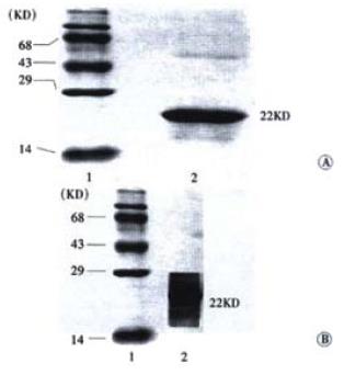
Figure 4 SDS-PAGE analysis and Western blot of endostatin expressed in supernatant of viral transduced SMMC7721 cells; A: SDS-PAGE analysis; 1, protein marker; 2, supernatant of SMMC7721 cells transfected with pLNCX-Endo; B: Western blot analysis; 1, protein marker; 2, supernatant of SMMC7721 cells transfected with pLNCX-Endo
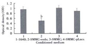
Figure 5 Inhibition of endothelial cell proliferation by conditioned medium from transfected and untransfected cells.
Conditioned medium from endostatin-transfected SMMC-Endo cells (2), conditioned medium from SMMC7721 cells (3), and conditioned medium from SMMC-pLncx cells (4) were concentrated and applied to cultivate with HUVEC cells grown in 40-well plate. Three days later, cell number, as measured by absorbance (OD), was then quantified by using a colorimetric MTT assay. Bars, SD. bP < 0.01, compared with conditioned medium from control SMMC-pLncx cells.
-
Citation: Wang X, Liu FK, Li X, Li JS, Xu GX. Inhibitory effect of endostatin expressed by human liver carcinoma SMMC7721 on endothelial cell proliferation
in vitro . World J Gastroenterol 2002; 8(2): 253-257 - URL: https://www.wjgnet.com/1007-9327/full/v8/i2/253.htm
- DOI: https://dx.doi.org/10.3748/wjg.v8.i2.253


