Copyright
©The Author(s) 2001.
World J Gastroenterol. Oct 15, 2001; 7(5): 678-684
Published online Oct 15, 2001. doi: 10.3748/wjg.v7.i5.678
Published online Oct 15, 2001. doi: 10.3748/wjg.v7.i5.678
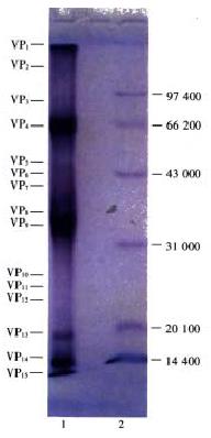
Figure 1 Protein polypetide map of NGVEV-CN virus.
1. Polypetide of NGVEV-CN virus; 2. Low MW. standard protein.
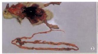
Figure 2 Particular coagulative embolus was formed in small in testine of death gosling (13 d postinfection), the length is over 40 cm.
Haemorrhage was occured in small intestine wall and be dyed red.
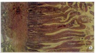
Figure 3 Many pieces of falled epidermal cells in duodenum cav ity (arrow 1), coagulative necroses was occured in some villus top (arrow 2).
(150 × , H.E)
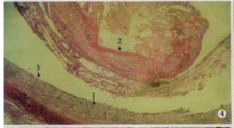
Figure 4 Particular fibrinous and necrosed enteritis of ileum: necrosed mucosae (arrow 1) and fibrinous edudate coagulated into artificial me mbrane and dropped into the cavity (arrow 2), and surface of separation boundry was smooth (arrow 3).
(100 × , H.E)
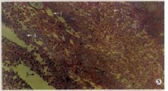
Figure 5 Particular fibrinous and necrosed enteritis of ileum: the embo lus consisted of the necrosed mucosal tissue coagulated and dropped materials, it include fibrinous edudate which like thread and inflammatory cells.
Fibrin (arrow 1), necrosed cells (arrow 2), inflammatory cells (arrow 3) and bacteria (arrow 4). (400 × , H.E)
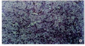
Figure 6 Fatty degeneration were occured in liver cells.
(500 × , H.E)
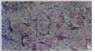
Figure 7 Kidney: Hyperaemia.
Granular degeneration was occured seriously in kidney small vessles (arrow 1), or even vacuolar degeneration (arrow 2). (500 × , H.E)
- Citation: Cheng AC, Wang MS, Chen XY, Guo YF, Liu ZY, Fang PF. Pathogenic and pathological characteristic of new type gosling viral enteritis first observed in China. World J Gastroenterol 2001; 7(5): 678-684
- URL: https://www.wjgnet.com/1007-9327/full/v7/i5/678.htm
- DOI: https://dx.doi.org/10.3748/wjg.v7.i5.678


