Copyright
©The Author(s) 2001.
World J Gastroenterol. Oct 15, 2001; 7(5): 678-684
Published online Oct 15, 2001. doi: 10.3748/wjg.v7.i5.678
Published online Oct 15, 2001. doi: 10.3748/wjg.v7.i5.678
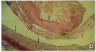
Figure 4 Particular fibrinous and necrosed enteritis of ileum: necrosed mucosae (arrow 1) and fibrinous edudate coagulated into artificial me mbrane and dropped into the cavity (arrow 2), and surface of separation boundry was smooth (arrow 3).
(100 × , H.E)
- Citation: Cheng AC, Wang MS, Chen XY, Guo YF, Liu ZY, Fang PF. Pathogenic and pathological characteristic of new type gosling viral enteritis first observed in China. World J Gastroenterol 2001; 7(5): 678-684
- URL: https://www.wjgnet.com/1007-9327/full/v7/i5/678.htm
- DOI: https://dx.doi.org/10.3748/wjg.v7.i5.678


