Copyright
©The Author(s) 2001.
World J Gastroenterol. Feb 15, 2001; 7(1): 98-101
Published online Feb 15, 2001. doi: 10.3748/wjg.v7.i1.98
Published online Feb 15, 2001. doi: 10.3748/wjg.v7.i1.98
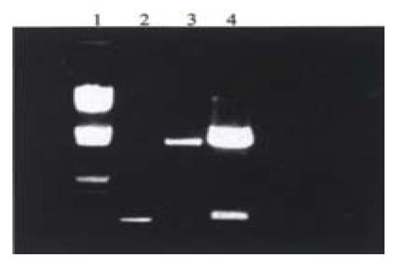
Figure 1 Analysis of recombinant plasmid by restriction endonuclease digestion.
1. λ DNA/Eco R I + Hind III; 2. PCR product; 3. pHTNS5/Bam H I + Kpn I; 4. pFHTNS5/Bam H I + Kpn I.
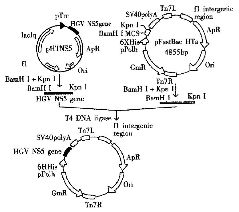
Figure 2 Construction of recombinant plasmid pFHTNS5.
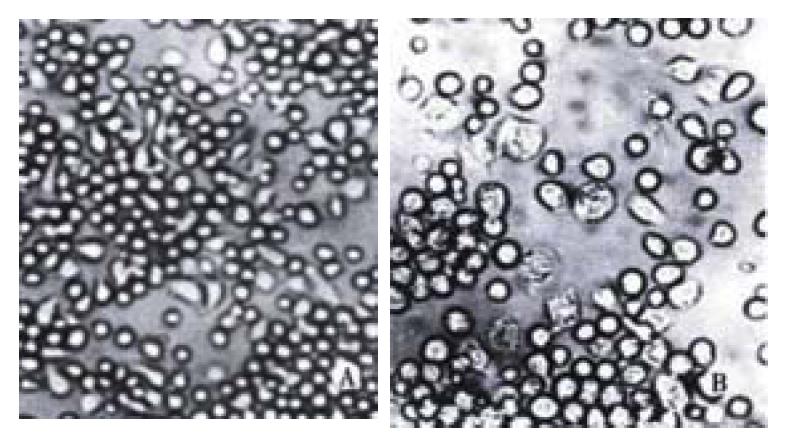
Figure 3 Morphology of uninfected, transfected and infected Sf9 cells (100 ×).
A: Uninfected cells; B: Transfected and infected cells
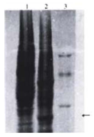
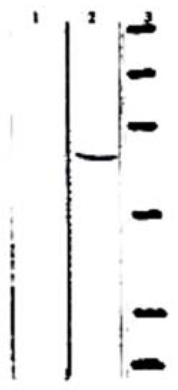
Figure 4 SDS-PAGE analysis of expressed HGV NS5 protein.
1. Uninfected sf9 cells; 2. sf9 cells infected with recombinant viruses; 3. Protein relative molecular mass standards. Arrow indicates the position of recombinant protein.
Figure 5 Western-blot analysis of recombinant protein HGV NS5.
1. Uninfected sf9 cells; 2. sf9 cells infected with recombinant viruses; 3. Protein relative molecular mass standards.
- Citation: Ren H, Zhu FL, Zhu SY, Song YB, Qi ZT. Immunogenicity of HGV NS5 protein expressed from Sf9 insect cells. World J Gastroenterol 2001; 7(1): 98-101
- URL: https://www.wjgnet.com/1007-9327/full/v7/i1/98.htm
- DOI: https://dx.doi.org/10.3748/wjg.v7.i1.98


