Copyright
©The Author(s) 2000.
World J Gastroenterol. Aug 15, 2000; 6(4): 588-592
Published online Aug 15, 2000. doi: 10.3748/wjg.v6.i4.588
Published online Aug 15, 2000. doi: 10.3748/wjg.v6.i4.588
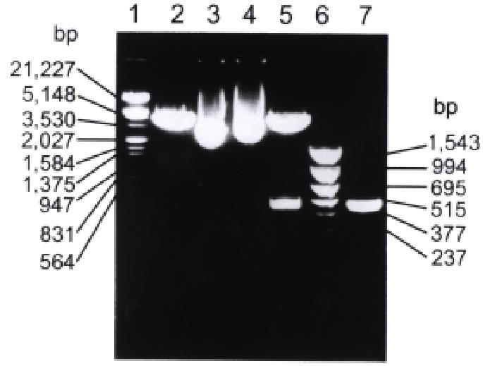
Figure 1 Restriction enzyme digestion and PCR analysis of recombinant plasmid pBV220/hCaM III.
1. λ DNA/Hind III+EcoR I marker; 2. pBV220 digested with EcoR I and BamH I; 3. pBV220; 4. pBV220/hCaM III; 5. pBV220/hCaM III digested with EcoR I and BamH I; 6. pBR322/Hinf I marker; 7. PCR product of hCaM III.
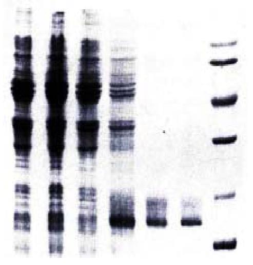
Figure 2 SDS-PAGE analysis of hCaM expression and purification.
1. Uninduced DH5α/pBV220; 2. Induced DH5α/pBV220; 3. Uninduced DH5α/pBV220-hCaM III; 4. Induced DH5α/pBV220-hCaM III; 5. Purified rhCaM by Phenyl-sepharose CL-4B column; 6. Standard Human brain CaM (Sigma); 7. Protein molecular weight marker.
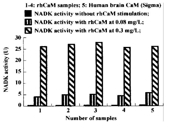
Figure 3 Activation of NADK by rhCaM.
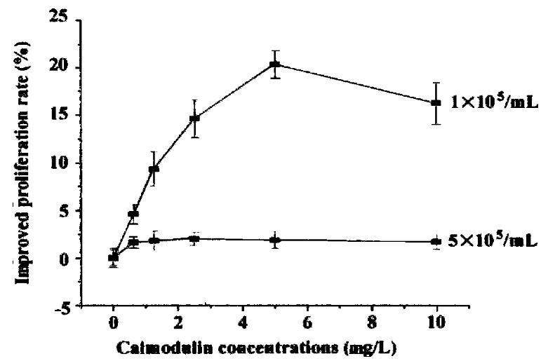
Figure 4 The effect of extracellular rhCaM on cultured SP2/0 cells.
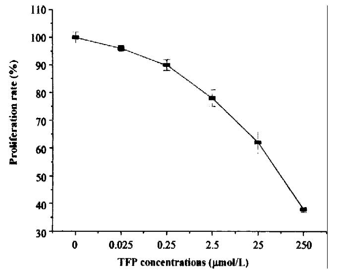
Figure 5 The inhibitory effect of CaM-antagonist TFP on cell proliferation.
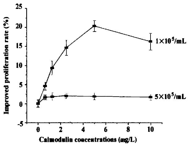
Figure 6 The effect of extracellular rhCaM on TFP inhibited cells.
- Citation: Li XJ, Wu JG, Si JL, Guo DW, Xu JP. High-level expression of human calmodulin in E.coli and its effects on cell proliferation. World J Gastroenterol 2000; 6(4): 588-592
- URL: https://www.wjgnet.com/1007-9327/full/v6/i4/588.htm
- DOI: https://dx.doi.org/10.3748/wjg.v6.i4.588


