Copyright
©The Author(s) 2000.
World J Gastroenterol. Feb 15, 2000; 6(1): 96-101
Published online Feb 15, 2000. doi: 10.3748/wjg.v6.i1.96
Published online Feb 15, 2000. doi: 10.3748/wjg.v6.i1.96
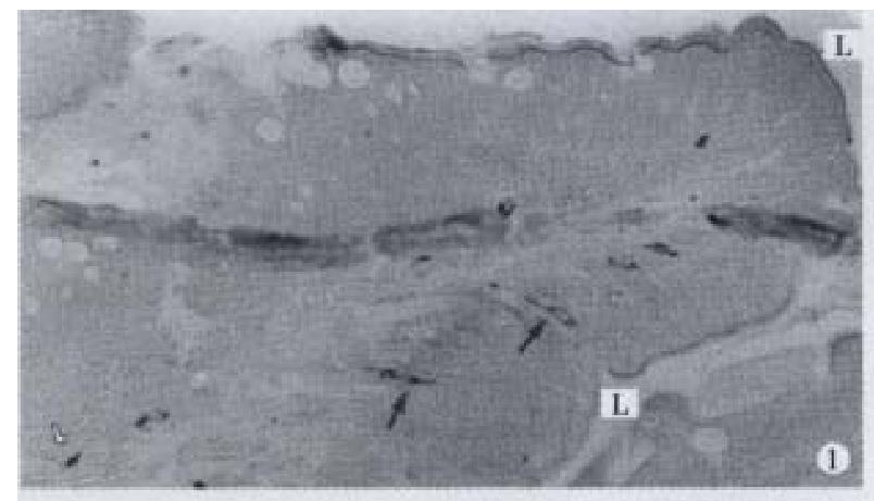
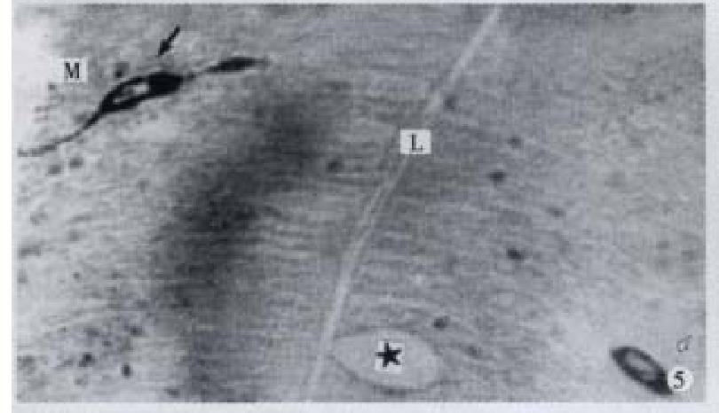
Figure 1 IR endocrine cells in epithelium (↑) and lamina propria (-), × 100
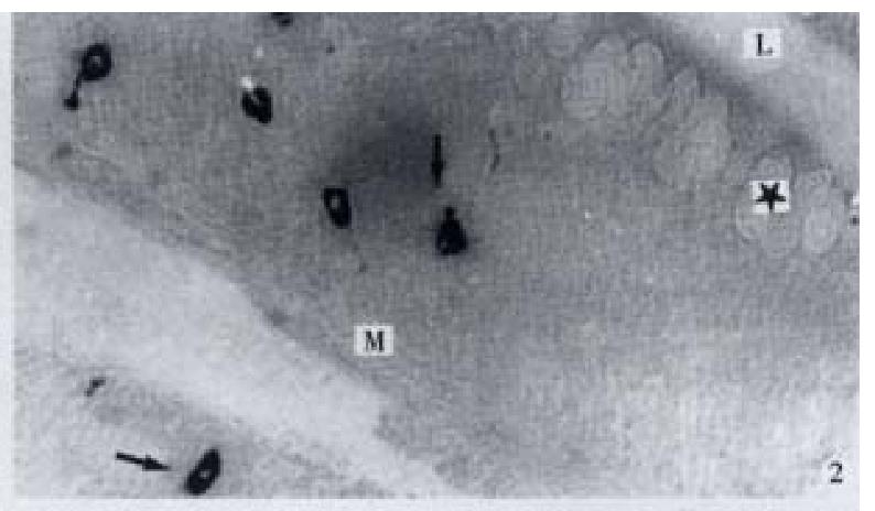
Figure 2 IR endocrine cells (↑) in bottom part of the gut f old, × 200
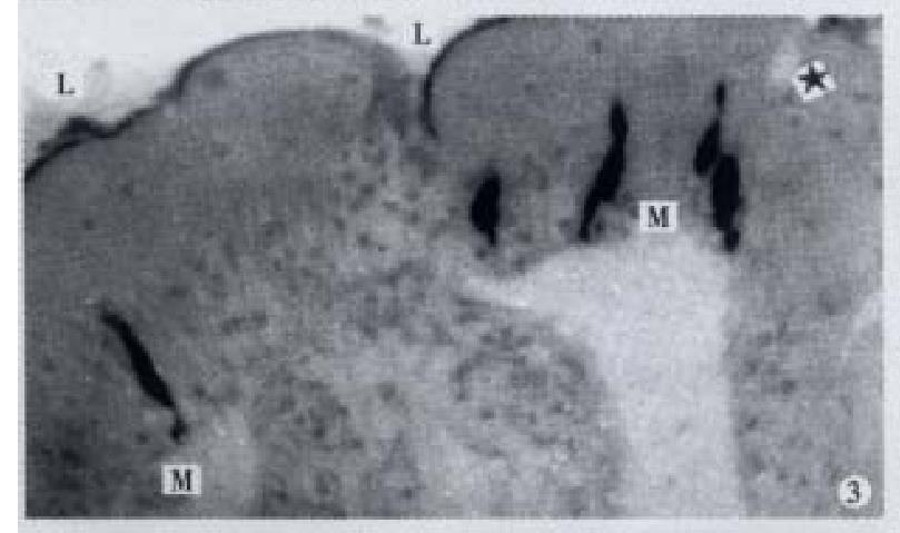
Figure 3 IR endocrine cells in apical part of the gut fold, × 400
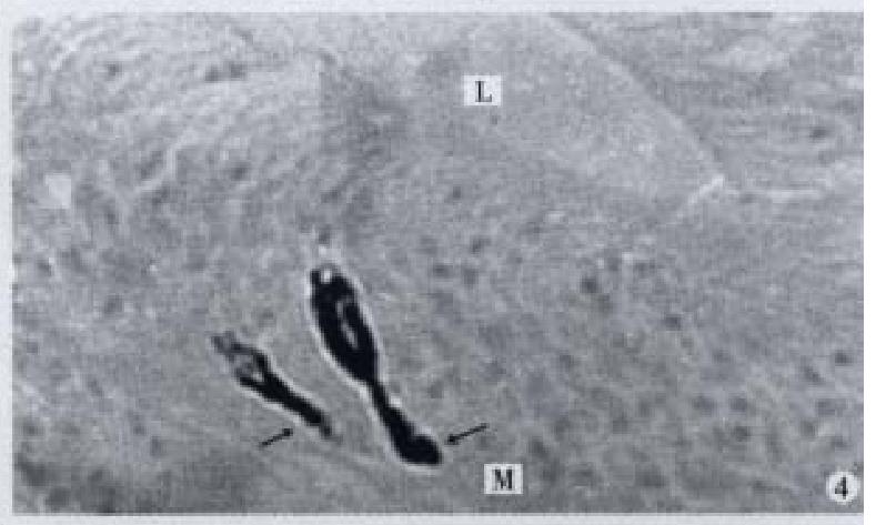
Figure 4 Basal processes (↑) extended to basement membrane, × 600
Figure 5 IR endocrine cells of open type (↑) and close type (-), × 600
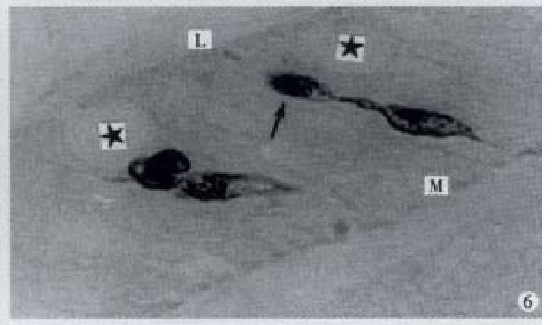
Figure 6 Enlarged process (↑) in sac-shaped, × 1000
Figure 7 Apical process (↑) as sac in shape, basal process (-) like synapse, × 1000
Figure 8 Apical process (↑) as sac in shape, basal process (-) like synapse, × 1000
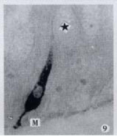
Figure 9 Basal process (-) extended to basement membrane, × 1000
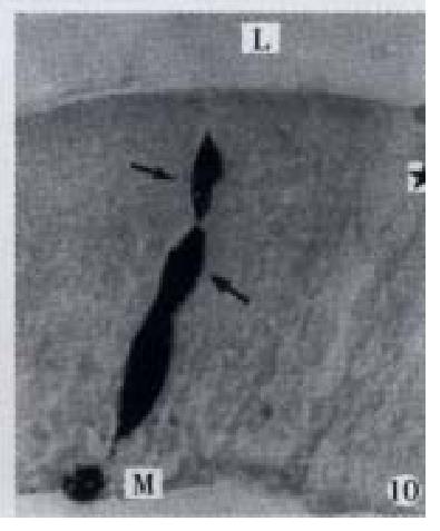
Figure 10 Apical process (↑) as sac in shape, basal process (-) formed an enlarged synapse-like structure, × 1000-In figures, ★: goblet cell; L: gut lumen; M: basement membrane.
- Citation: Pan QS, Fang ZP, Zhao YX. Immunocytochemical identification and localization of APUD cells in the gut of seven stomachless teleost fishes. World J Gastroenterol 2000; 6(1): 96-101
- URL: https://www.wjgnet.com/1007-9327/full/v6/i1/96.htm
- DOI: https://dx.doi.org/10.3748/wjg.v6.i1.96


