Copyright
©The Author(s) 2000.
World J Gastroenterol. Feb 15, 2000; 6(1): 57-60
Published online Feb 15, 2000. doi: 10.3748/wjg.v6.i1.57
Published online Feb 15, 2000. doi: 10.3748/wjg.v6.i1.57
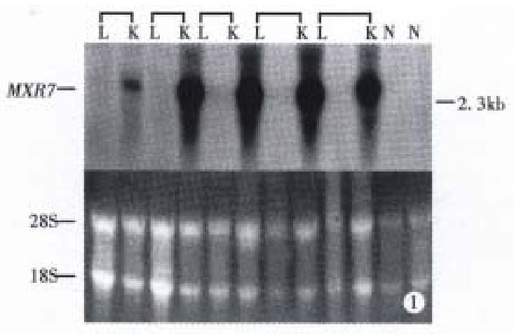
Figure 1 Northern blot analysis of MXR7 in human HCC, paracancerous hepatic and normal liver tissues.
L: paratumor tissue; K: hepatoma tissue; N: normal liver tissue. 28S and 18S rRNAs were used for evalua ting the quality and quantity of RNA loading.
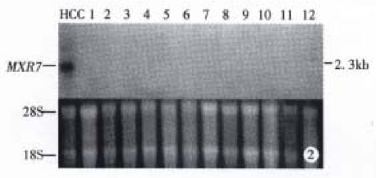
Figure 2 Northern blot analysis of MXR7 in 12 different human normal tissues.
Lane 1: liver; Lane 2: lung; Lane 3: kidney; Lane 4: heart; Lane 5: brain; Lane 6: small intestine; Lane 7: colon; Lane 8: testis; Lane 9: spleen; Lane 10: stomach; Lane 11: cyst; Lane 12: pancreas; HCC as a positive control. 28S and 18S rRNAs were used for evaluating the qualit y and quantity of RNA loading.
Figure 3 Northern blot analysis of MXR7 in 7 non-liver tumor tissues.
Lane 1: gastric adenocarcinoma; Lane 2: sigmoid adenocarc inoma; Lane 3: gastric adenocarcinoma; Lane 4: left colon-ileal malignant mesot helioma; Lane 5: gastric adenocarcinoma; Lane 6: uterus adenomyoma; Lane 7: coli c familial polyadenomatosis; HCC as a positive control. 28S and 18S rRNAs were used for evaluating the quality and quantity of RNA loading.
- Citation: Zhou XP, Wang HY, Yang GS, Chen ZJ, Li BA, Wu MC. Cloning and expression of MXR7 gene in human HCC tissue. World J Gastroenterol 2000; 6(1): 57-60
- URL: https://www.wjgnet.com/1007-9327/full/v6/i1/57.htm
- DOI: https://dx.doi.org/10.3748/wjg.v6.i1.57


