Copyright
©The Author(s) 1998.
World J Gastroenterol. Dec 15, 1998; 4(6): 513-515
Published online Dec 15, 1998. doi: 10.3748/wjg.v4.i6.513
Published online Dec 15, 1998. doi: 10.3748/wjg.v4.i6.513
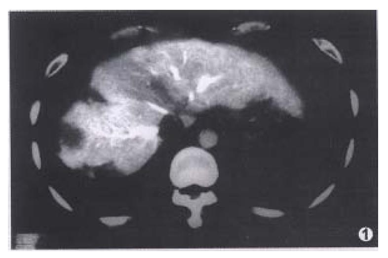
Figure 1 CTAP image obtained in a 42-year-old man with recurrence of HCC shows two tumors in right lobe.
The peripheral wedge-shaped perfusion defect is non-pathologic perfusion defect, confirmed by biopsy.
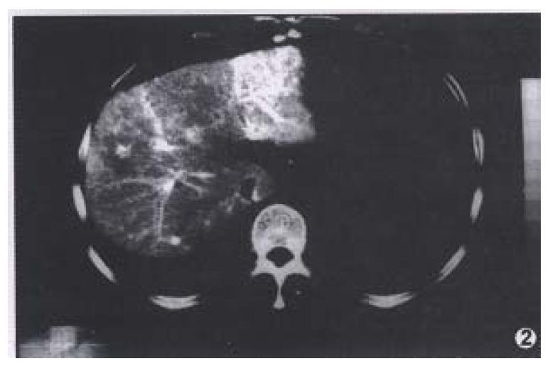
Figure 2 CTHA image obtained in a 45-year-old man shows a small round peripheral enhancement in right lobe which is non-pathologic enhancement.
No tumor is found in the operative pathology.
- Citation: Li L, Wu PH, Lin HG, Li JQ, Mo YX, Zheng L, Lu LX, Ruan CM, Chen L. Findings of non-pathologic perfusion defects by CT arterial portography and non-pathologic enhancement of CT hepatic arteriography. World J Gastroenterol 1998; 4(6): 513-515
- URL: https://www.wjgnet.com/1007-9327/full/v4/i6/513.htm
- DOI: https://dx.doi.org/10.3748/wjg.v4.i6.513


