Copyright
©The Author(s) 1998.
World J Gastroenterol. Feb 15, 1998; 4(1): 19-23
Published online Feb 15, 1998. doi: 10.3748/wjg.v4.i1.19
Published online Feb 15, 1998. doi: 10.3748/wjg.v4.i1.19
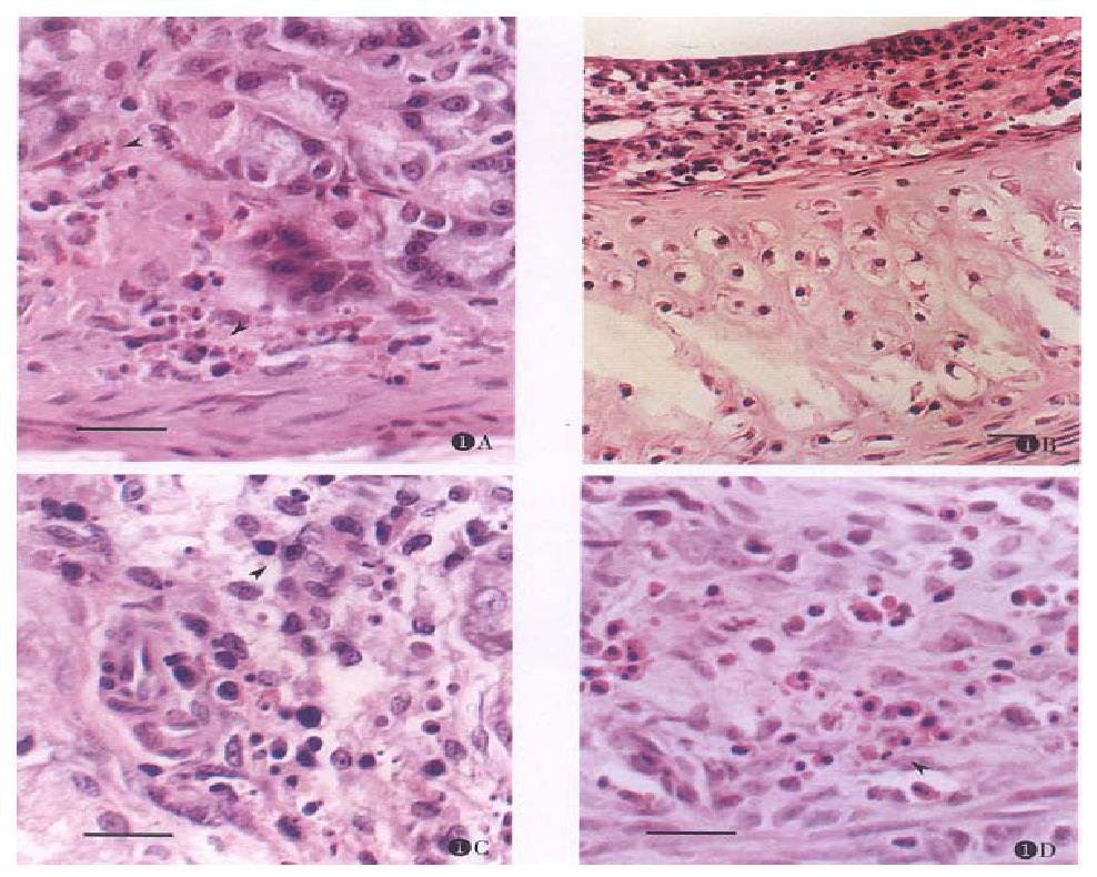
Figure 1 Occurrence of apoptosis in mucosa 3 h after injection of CHX (3.
0 mg·kg-1) (bar = 20.0 μm, H&E). (A) Stomach apoptotic cells (bodies) lie in the lamina propria (arrows) of the mucosa. The parietal cells, chief cells and enterochromaffin-like cells seem not to be affected by treatment of CHX. (D) and (C) apoptosis in lamina propria of uterus and lacrimal gland. The apoptotic cells (bodies) were phagocytised by macrophages. (B) Mucosa apoptosis in trachea.
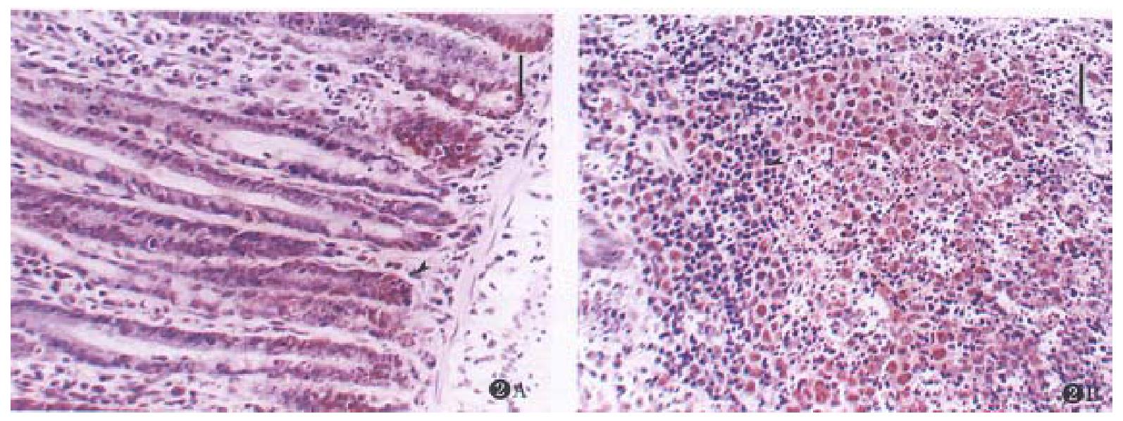
Figure 2 Expression of PCNA in mucosa after treated with CHX (3.
0 mg·kg-1) in 3 h. (bar = 20.0 μm, stained with hematoxylin). (A) Colon. The apoptosis of crypt epithelial cells on the basement of mucosa were seen with expression of PCNA (arrow). (B) Peyer’s patches in ileum. Apoptotic cells around the germinal center with expression of PCNA. The lymphocytes in parafollicular T-cell zone were morphologically normal and were negatively expressed with PCNA (arrow).
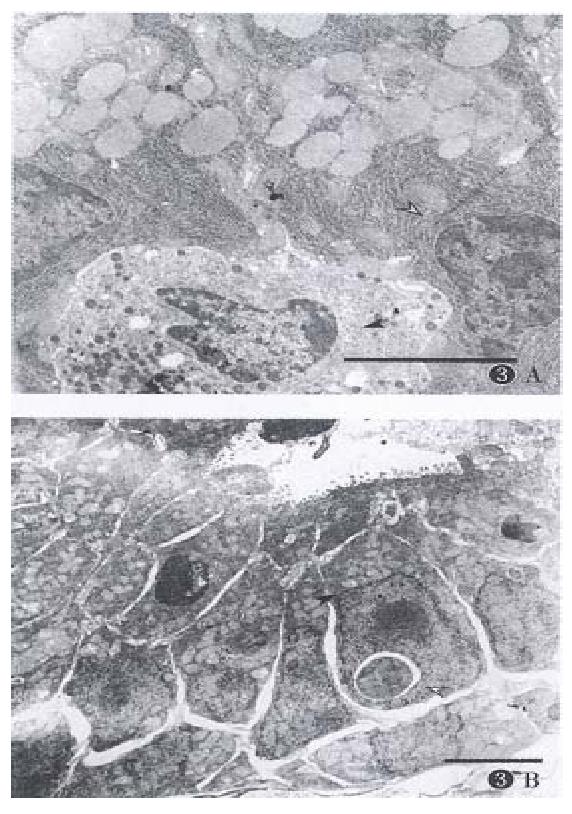
Figure 3 Morphological changes of cells after given CHX (3.
0 mg·kg-1) 3 h later (bar = 4.0 μm). (A) Stomach. Normal appearance of chief cell (hollow arrow) and ECL cell (black arrow). (B) Jejunum. Condensed nucleus (hollow arrow) and proliferated mitochondria (black arrow).
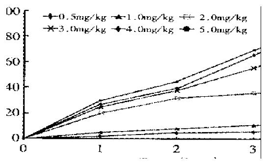
Figure 4 The time- and dosage-dependent effect of CHX on apoptotic bodies (cells) formation in rat uterus.
- Citation: Chen XQ, Zhang WD, Song YG, Zhou DY. Induction of apoptosis of lymphocytes in rat mucosal immune system. World J Gastroenterol 1998; 4(1): 19-23
- URL: https://www.wjgnet.com/1007-9327/full/v4/i1/19.htm
- DOI: https://dx.doi.org/10.3748/wjg.v4.i1.19


