
















©The Author(s) 1998.
World J Gastroenterol. Feb 15, 1998; 4(1): 19-23
Published online Feb 15, 1998. doi: 10.3748/wjg.v4.i1.19
Published online Feb 15, 1998. doi: 10.3748/wjg.v4.i1.19
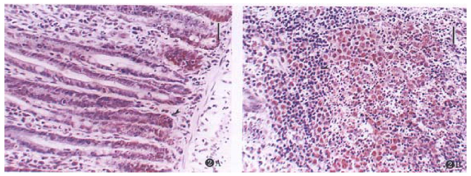
Figure 2 Expression of PCNA in mucosa after treated with CHX (3.
0 mg·kg-1) in 3 h. (bar = 20.0 μm, stained with hematoxylin). (A) Colon. The apoptosis of crypt epithelial cells on the basement of mucosa were seen with expression of PCNA (arrow). (B) Peyer’s patches in ileum. Apoptotic cells around the germinal center with expression of PCNA. The lymphocytes in parafollicular T-cell zone were morphologically normal and were negatively expressed with PCNA (arrow).
- Citation: Chen XQ, Zhang WD, Song YG, Zhou DY. Induction of apoptosis of lymphocytes in rat mucosal immune system. World J Gastroenterol 1998; 4(1): 19-23
- URL: https://www.wjgnet.com/1007-9327/full/v4/i1/19.htm
- DOI: https://dx.doi.org/10.3748/wjg.v4.i1.19