Copyright
©The Author(s) 1998.
World J Gastroenterol. Feb 15, 1998; 4(1): 19-23
Published online Feb 15, 1998. doi: 10.3748/wjg.v4.i1.19
Published online Feb 15, 1998. doi: 10.3748/wjg.v4.i1.19
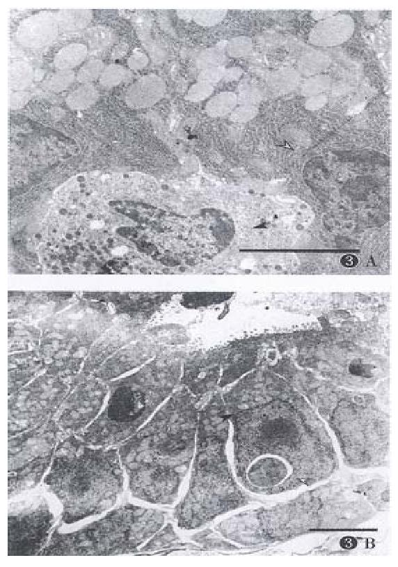
Figure 3 Morphological changes of cells after given CHX (3.
0 mg·kg-1) 3 h later (bar = 4.0 μm). (A) Stomach. Normal appearance of chief cell (hollow arrow) and ECL cell (black arrow). (B) Jejunum. Condensed nucleus (hollow arrow) and proliferated mitochondria (black arrow).
- Citation: Chen XQ, Zhang WD, Song YG, Zhou DY. Induction of apoptosis of lymphocytes in rat mucosal immune system. World J Gastroenterol 1998; 4(1): 19-23
- URL: https://www.wjgnet.com/1007-9327/full/v4/i1/19.htm
- DOI: https://dx.doi.org/10.3748/wjg.v4.i1.19


