Copyright
©The Author(s) 2025.
World J Gastroenterol. May 14, 2025; 31(18): 103778
Published online May 14, 2025. doi: 10.3748/wjg.v31.i18.103778
Published online May 14, 2025. doi: 10.3748/wjg.v31.i18.103778
Figure 1 Intestinal infarction specimens and microscopic findings.
A: Necrotic bowel; B: Only the tissue structure can be seen in the muscular layer [hematoxylin & eosin (H&E) 100 ×]; C: The mucosa adjacent to the necrotic bowel exhibited lymphocytic and plasmacytic infiltration, with focal chronic enteritis and approximately 10 eosinophils per high-power field (H&E 20 ×).
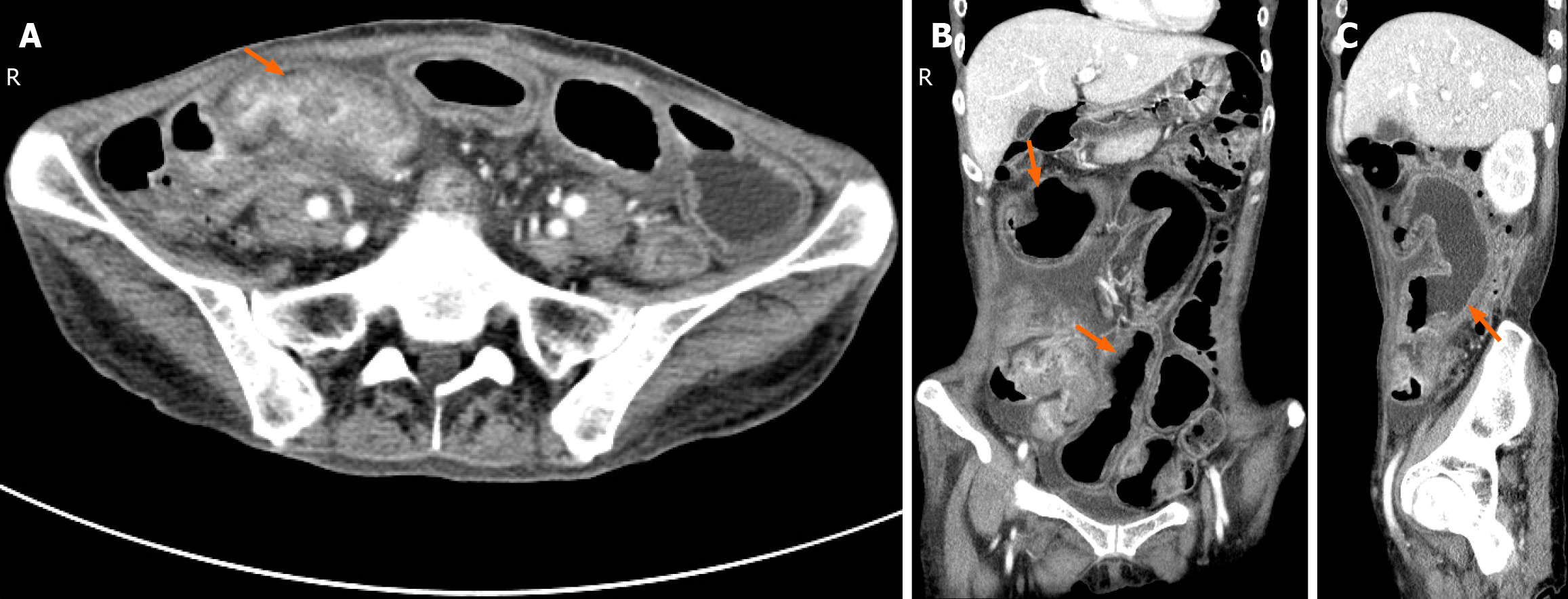
Figure 2 Abdominal enhanced computed tomography image.
A: Horizontal plane, Thickening of the intestinal wall of the terminal ileum, narrowing of the lumen, localized peritoneal thickening and accumulation of intestinal tubes, and enhancement of the intestinal wall (arrow); B: Coronal plane; C: Right sagittal plane, Proximal small bowel dilation, multiple bowel wall thickening and layered enhancement (arrows).
Figure 3 Endoscopic and specimen images.
A and B: Terminal ileum, approximately 8 cm long longitudinal ulcer, bleeding on the mucosal surface, surrounding small granular hyperplasia; C: Surgical image, tortuous and lumpy terminal ileum (triangle), dilated and edematous proximal small bowel (arrow); D: Resected bowel, intestinal wall thickening with a constricted ring (triangle) and a longitudinal ulcer (arrow) in the adjacent area.
Figure 4 Microscopic observation of lesions.
A: Small intestinal villous atrophy with extensive pyloric gland metaplasia, submucosal fibrous tissue proliferation, and infiltration of lymphocytes, plasma cells, and granulocytes in both the mucosa and submucosa, with focal abscess formation and approximately 15 eosinophils per high-power field. No typical granulomatous lesions were observed on the slide [hematoxylin & eosin (H&E) 20 ×]; B: Irregular proliferation of smooth muscle cells in mesenteric veins, eccentric hyperplasia (H&E 100 ×); C: Mesenteric vein intimal hyperplasia (arrow), lumen stenosis, no thrombus, and no obvious abnormality in the accompanying artery (triangle) (H&E 100 ×).
- Citation: Jiang ZX, Yuan LW, Peng LX, Yang LC, Zhang YW, Wu Q, Yao BJ, Wang XH. Idiopathic myointimal hyperplasia of the mesenteric veins affecting the small intestine alone: A case report and review of literature. World J Gastroenterol 2025; 31(18): 103778
- URL: https://www.wjgnet.com/1007-9327/full/v31/i18/103778.htm
- DOI: https://dx.doi.org/10.3748/wjg.v31.i18.103778


