Copyright
©The Author(s) 2025.
World J Gastroenterol. May 14, 2025; 31(18): 103778
Published online May 14, 2025. doi: 10.3748/wjg.v31.i18.103778
Published online May 14, 2025. doi: 10.3748/wjg.v31.i18.103778
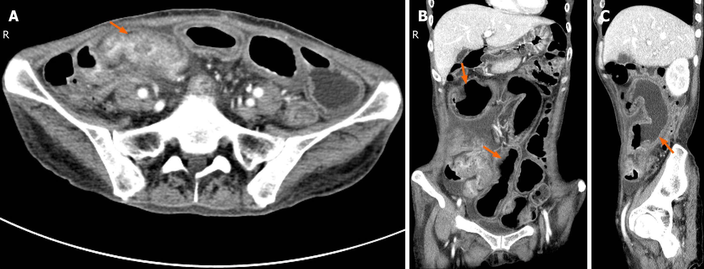
Figure 2 Abdominal enhanced computed tomography image.
A: Horizontal plane, Thickening of the intestinal wall of the terminal ileum, narrowing of the lumen, localized peritoneal thickening and accumulation of intestinal tubes, and enhancement of the intestinal wall (arrow); B: Coronal plane; C: Right sagittal plane, Proximal small bowel dilation, multiple bowel wall thickening and layered enhancement (arrows).
- Citation: Jiang ZX, Yuan LW, Peng LX, Yang LC, Zhang YW, Wu Q, Yao BJ, Wang XH. Idiopathic myointimal hyperplasia of the mesenteric veins affecting the small intestine alone: A case report and review of literature. World J Gastroenterol 2025; 31(18): 103778
- URL: https://www.wjgnet.com/1007-9327/full/v31/i18/103778.htm
- DOI: https://dx.doi.org/10.3748/wjg.v31.i18.103778


