Copyright
©The Author(s) 2024.
World J Gastroenterol. Feb 7, 2024; 30(5): 499-508
Published online Feb 7, 2024. doi: 10.3748/wjg.v30.i5.499
Published online Feb 7, 2024. doi: 10.3748/wjg.v30.i5.499
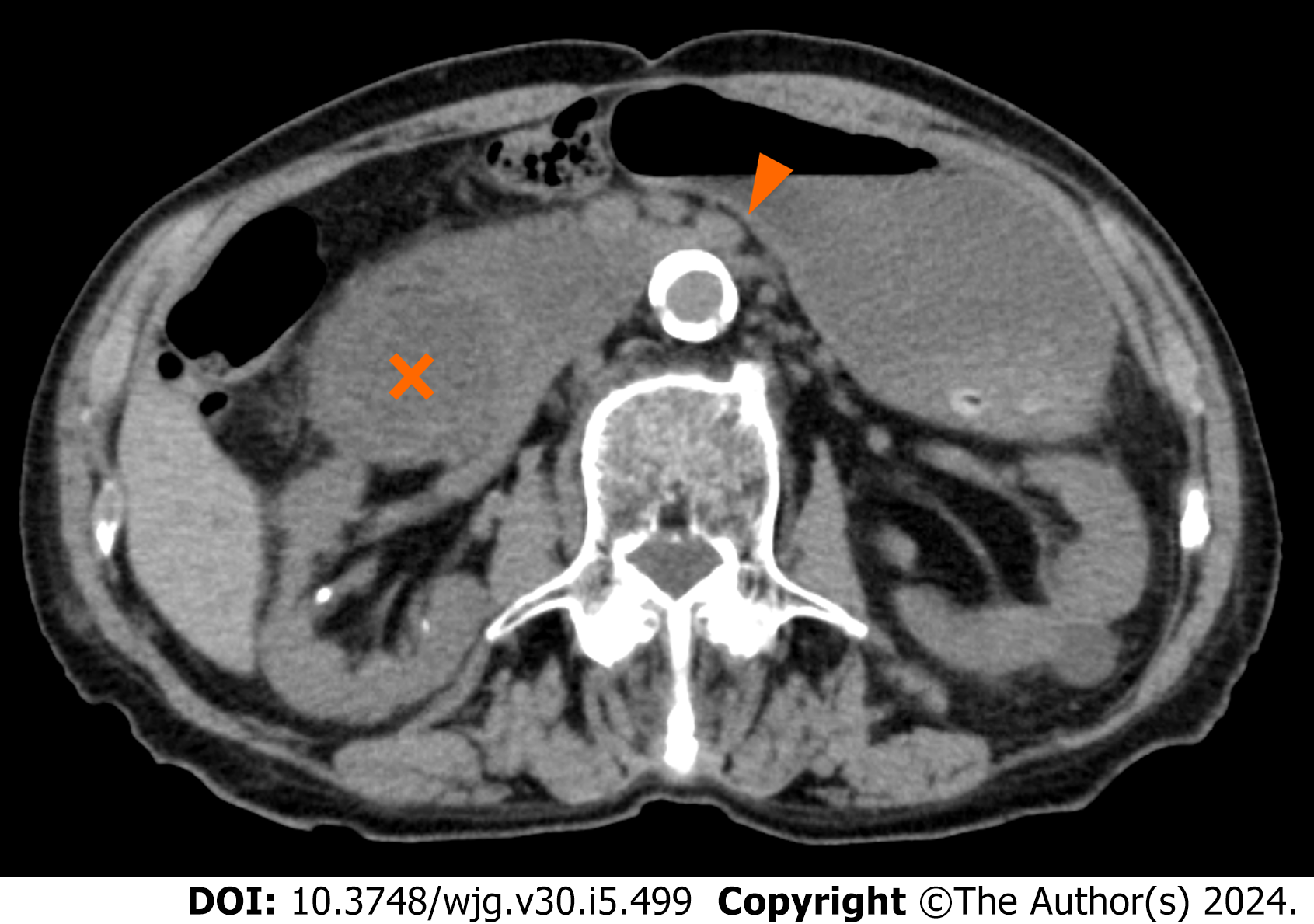
Figure 1 Computed tomography images of case 1.
The axial view of computed tomography showed the compression of the third portion of the duodenum between the superior mesenteric artery and the aorta (arrowhead) as well as markedly dilated duodenum (cross) and stomach.
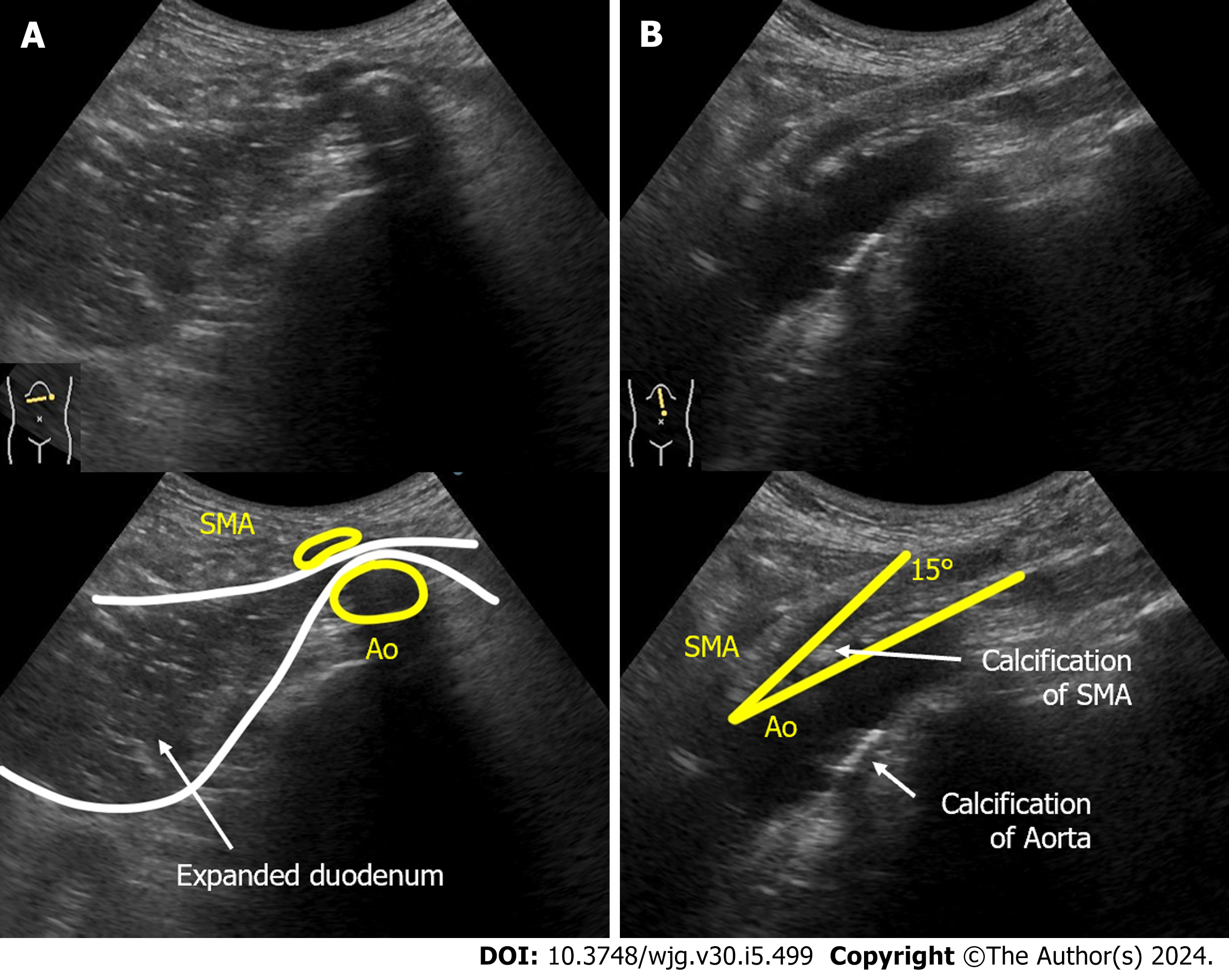
Figure 2 Abdominal ultrasonography images of case 1.
A: The supine position of ultrasonography epigastric transverse scan confirmed expansion of the third part of the duodenal lumen and extrinsic compression of the duodenum by the superior mesenteric artery (SMA). The distance between the SMA and the aorta (Ao) was narrowed to 4 mm; B: Epigastric longitudinal scan showed that the angle of SMA-Ao decreased to 15°. Calcifications (arteriosclerosis) of the aorta and the root of SMA was also observed. SMA: Superior mesenteric artery; Ao: Aorta.
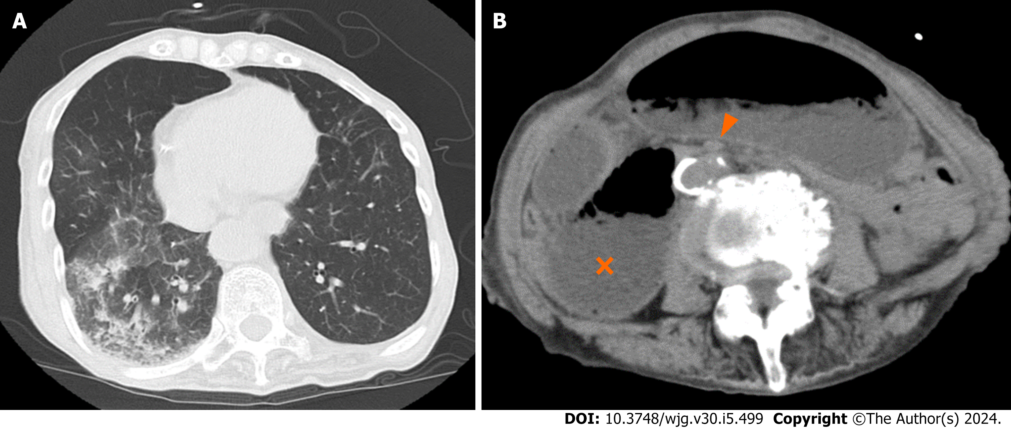
Figure 3 Computed tomography images of case 2.
A: The axial view of thoracic computed tomography (CT) indicated aspiration pneumonia; B: The axial view of abdominal CT demonstrates extensive dilatation of stomach and proximal duodenum (cross), between the superior mesenteric artery (arrow head) and the aorta.
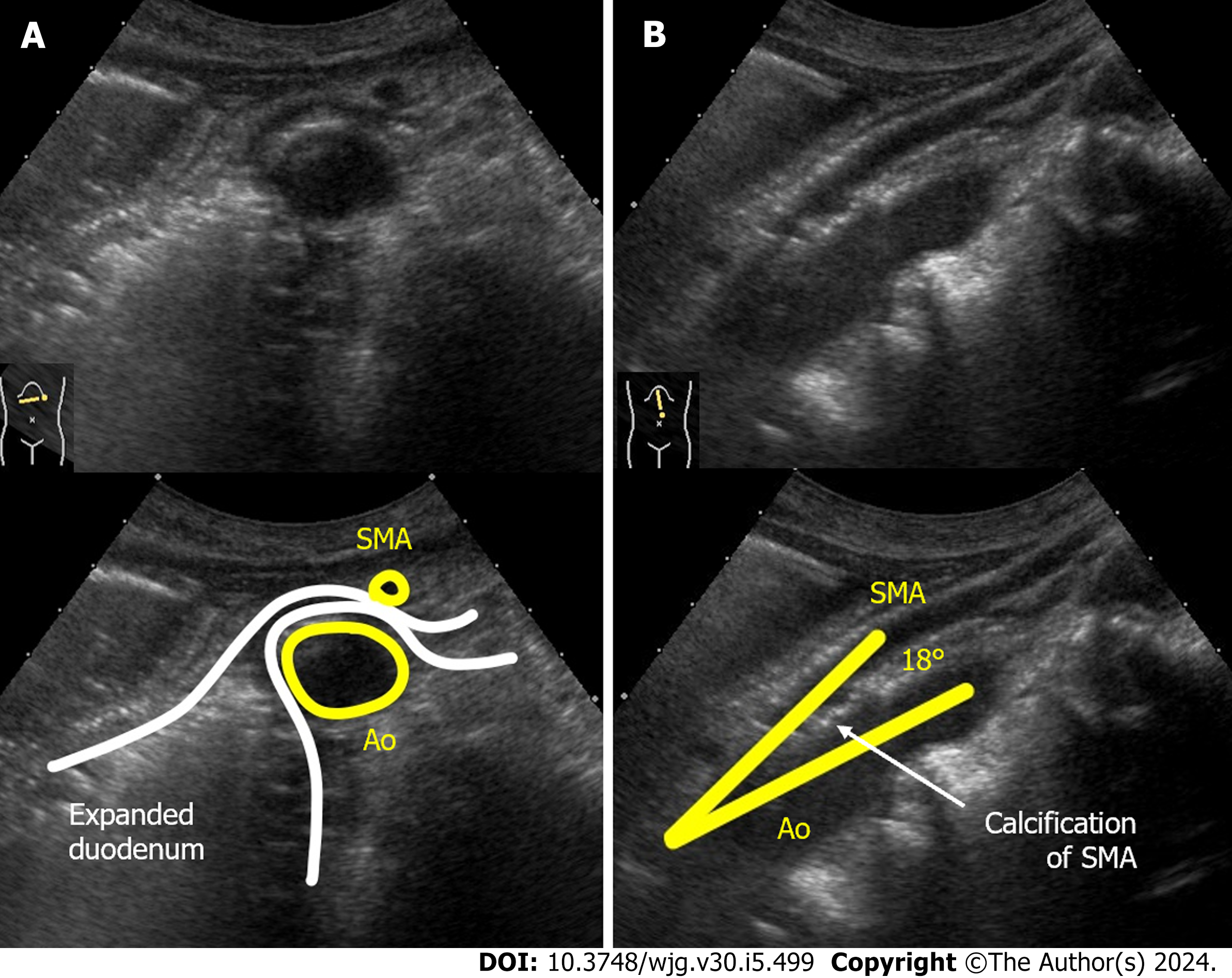
Figure 4 Dynamic ultrasonography images of case 2.
A: The supine position of ultrasonography confirmed expansion of the third part of the duodenal lumen and extrinsic compression of the duodenum by the superior mesenteric artery (SMA). The distance between the SMA and the aorta (Ao) was narrowed to 5 mm; B: Epigastric longitudinal scan showed that the angle of SMA-Ao decreased to 18°. Calcifications (arteriosclerosis) of the aorta and the root of SMA was also observed. SMA: Superior mesenteric artery; Ao: Aorta.
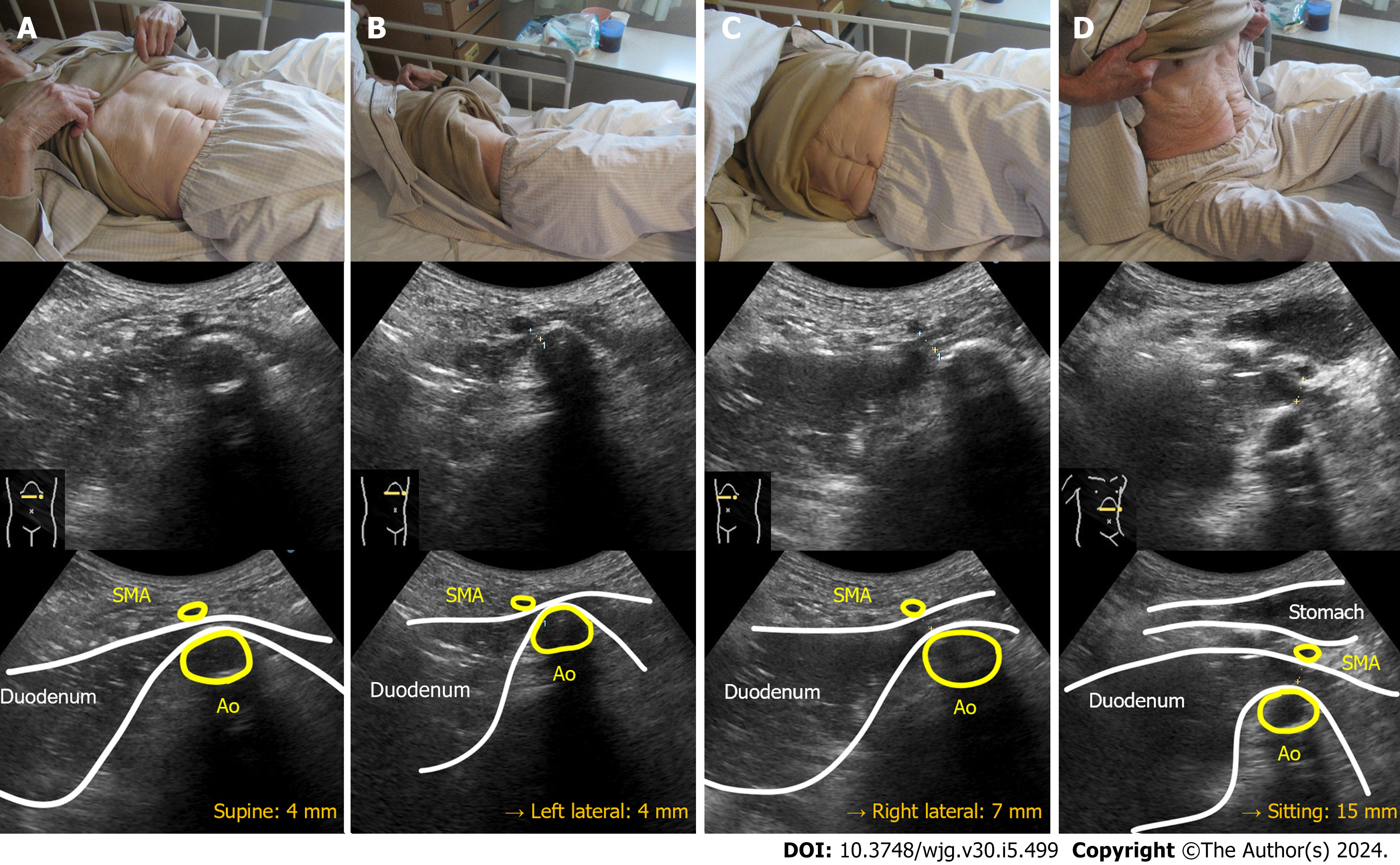
Figure 5 Dynamic ultrasonography images of case 1.
A: Narrowed superior mesenteric artery-aorta (SMA-Ao) distance (4 mm); B: Left lateral position did not improve the SMA-Ao distance (4 mm); C: Right lateral position increased the SMA-Ao distance to 7 mm and the duodenal gas could pass through; D: Sitting position (Fowler’s position) dramatically improved the SMA-Ao distance to 15 mm and the passage of duodenal contents. SMA: Superior mesenteric artery; Ao: Aorta.
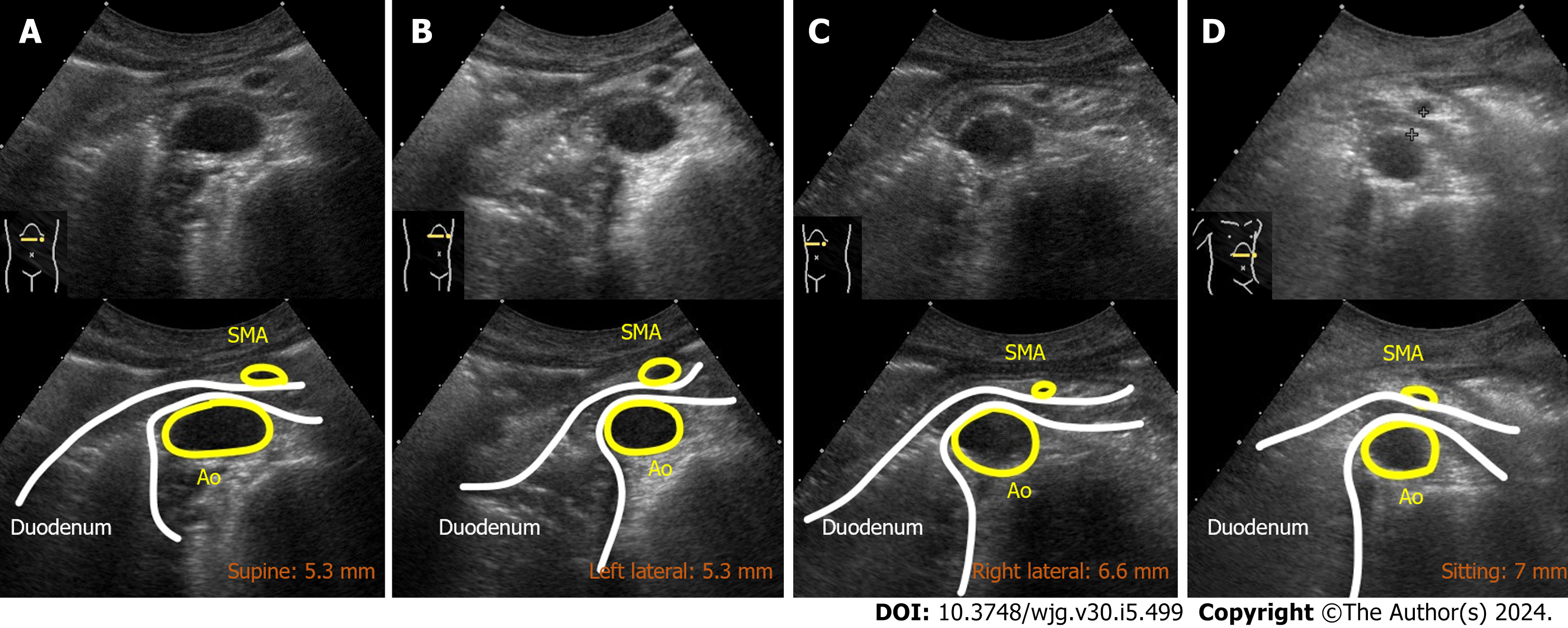
Figure 6 Dynamic ultrasonography images of case 2.
A: Supine position showed the narrowed superior mesenteric artery-aorta (SMA-Ao) distance (5.3 mm); B: Left lateral position did not improve the SMA-Ao distance (5.3 mm); C and D: Right lateral position and sitting position increased the SMA-Ao distance to 6.6 and 7.0 mm, respectively. SMA: Superior mesenteric artery; Ao: Aorta.
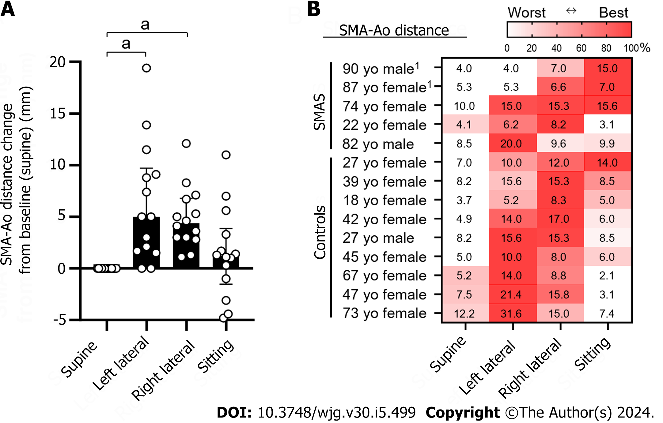
Figure 7 Superior mesenteric artery-aorta distance in four body positions in eight individuals.
A: Distance change of superior mesenteric artery-aorta (SMA-Ao) from the baseline position (supine position). Dots indicate individual results. Bars indicate median with interquartile range; B: Fourteen individuals included five cases of superior mesenteric artery syndrome and nine healthy controls (age, sex, and absolute distance of SMA-Ao mm). The absolute distance of SMA-Ao (mm) in thirteen individuals were shown in each position. The SMA-Ao distances were measured by ultrasonography. If it was difficult to detect the duodenum, the subjects ingested 200 mL of water which is a modified method of drinking-ultrasonography test. Statistical analysis was performed by GraphPad Prism version 10.0.2 with Dunn’s multiple comparisons tests with ANOVA. 1Indicates the present case. aP < 0.01. The heat map indicates the best and worst positions of the SMA-Ao distance in each individual. SMA: Superior mesenteric artery; Ao: Aorta; SMAS: Superior mesenteric artery syndrome.
- Citation: Hasegawa N, Oka A, Awoniyi M, Yoshida Y, Tobita H, Ishimura N, Ishihara S. Dynamic ultrasonography for optimizing treatment position in superior mesenteric artery syndrome: Two case reports and review of literature. World J Gastroenterol 2024; 30(5): 499-508
- URL: https://www.wjgnet.com/1007-9327/full/v30/i5/499.htm
- DOI: https://dx.doi.org/10.3748/wjg.v30.i5.499


