Copyright
©The Author(s) 2021.
World J Gastroenterol. Feb 14, 2021; 27(6): 470-486
Published online Feb 14, 2021. doi: 10.3748/wjg.v27.i6.470
Published online Feb 14, 2021. doi: 10.3748/wjg.v27.i6.470
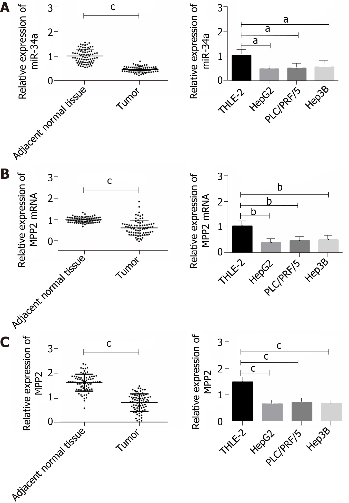
Figure 1 Low expression of microRNA 34a and membrane palmitoylated proteins in liver cancer.
A: Low expression of microRNA 34a (miR-34a) in liver cancer tissues and cells; B: The mRNA expression of membrane palmitoylated protein (MPP2) was low in liver cancer tissues and cells; and C: The protein expression of MMP2 in liver cancer tissues and cells; a indicates aP < 0.05, bP < 0.01, cP < 0.001).
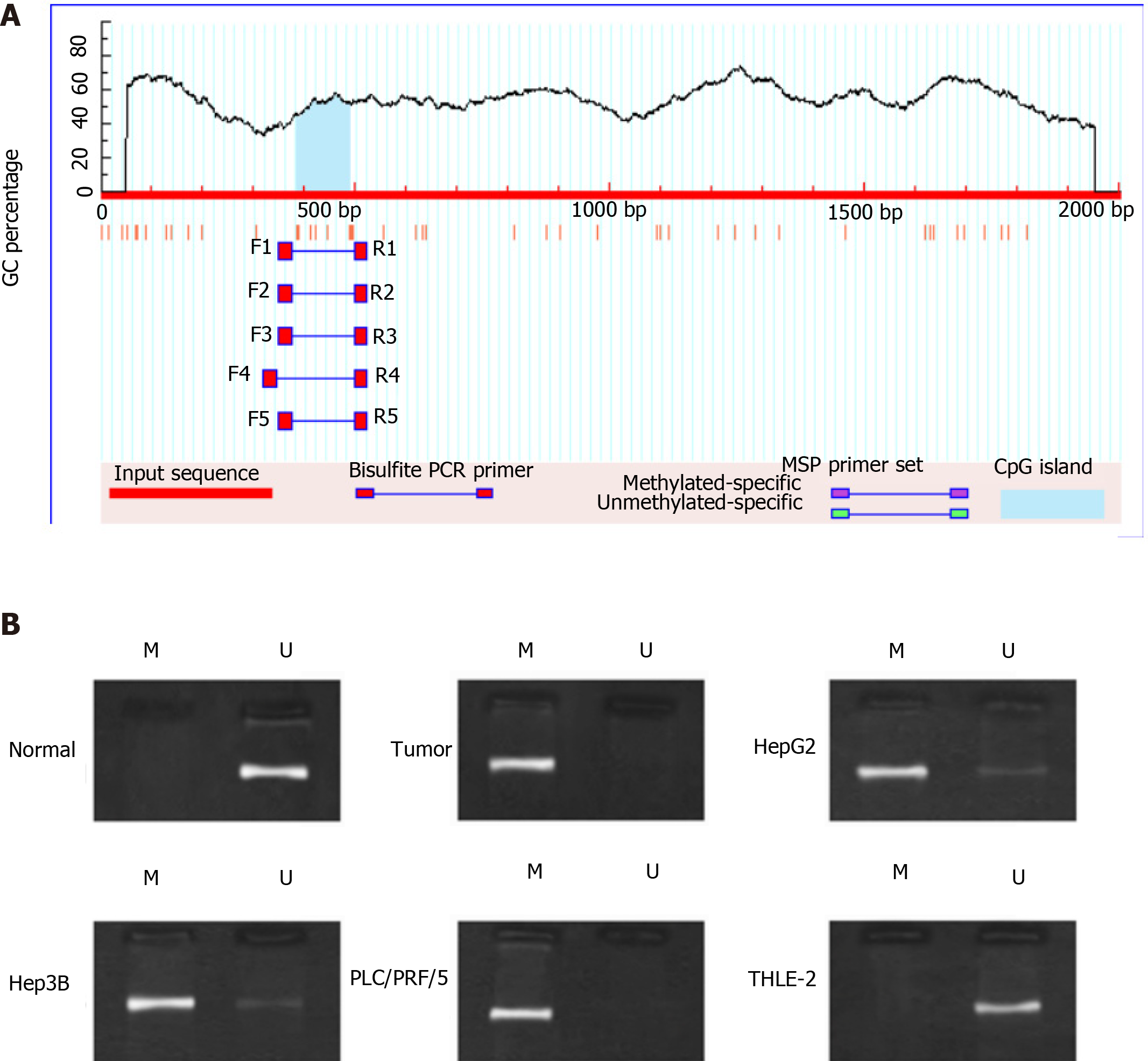
Figure 2 Highly methylated miR-34a in liver cancer.
A: Methylation site prediction; B: Methylation of miR-34a in liver cancer. MSP: Methylation-specific polymerase chain reaction.
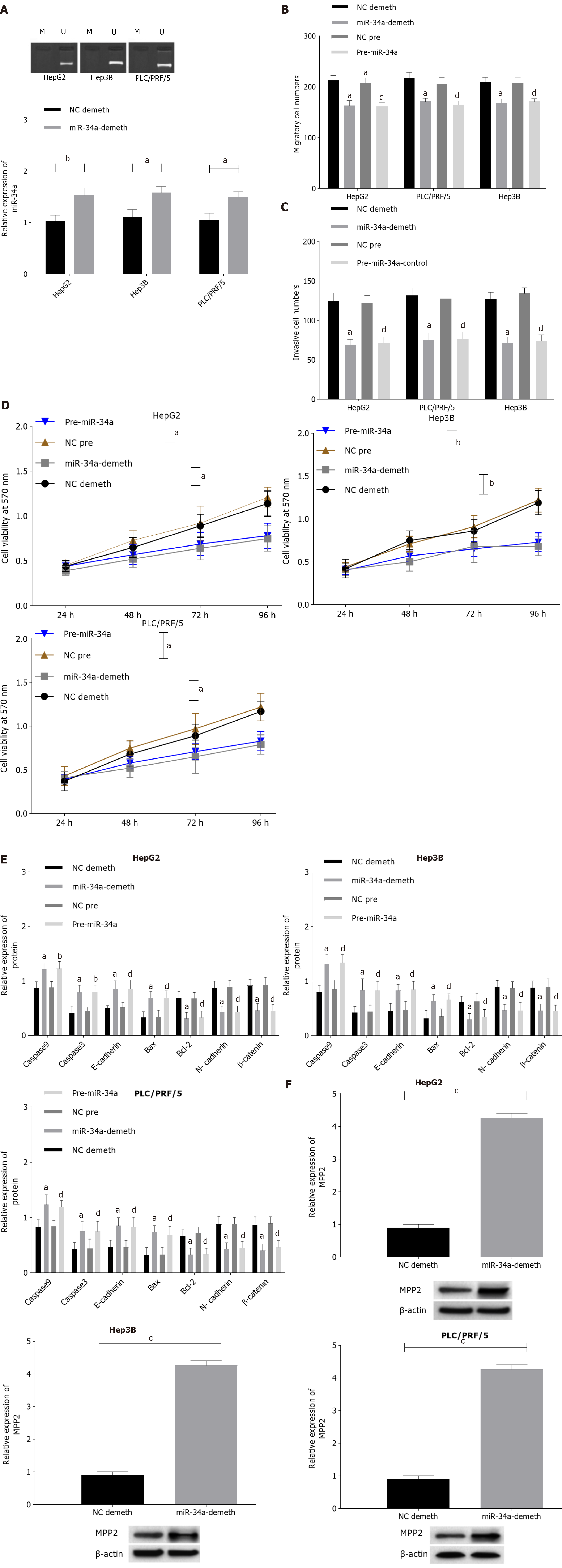
Figure 3 Upregulation of microRNA 34a promotes cell apoptosis and membrane palmitoylated protein expression, and inhibits cell proliferation, invasion, and migration.
A: Image of microRNA 34a (miR-34a) demethylation results; B: Upregulation of miR-34a inhibits cell migration; a indicates that compared with negative control (NC) demethylation group, aP < 0.05; bP < 0.01, and d indicates compared with the NC pre group, dP < 0.05; C: miR-34a upregulated the number of cell invasions; a indicates compared with the NC demethylation group, aP < 0.05; d indicates compared with the NC pre group, dP < 0.05; D: miR-34a upregulated and inhibited cell activity; a indicates aP < 0.05, bP < 0.01; E: Upregulation of miR-34a inhibited the downregulation of B-cell lymphoma 2 (Bcl-2), N- cadherin and β-catenin expression, while promoting caspase 9, caspase 3, E-cadherin and Bcl-2-associated X protein; a indicates compared with the NC demethylation group, aP < 0.05; and d indicates compared with the NC pre group, dP < 0.05; and F: Upregulation of miR-34a promoted the expression of membrane palmitoylated proteins, c indicates cP < 0.001.
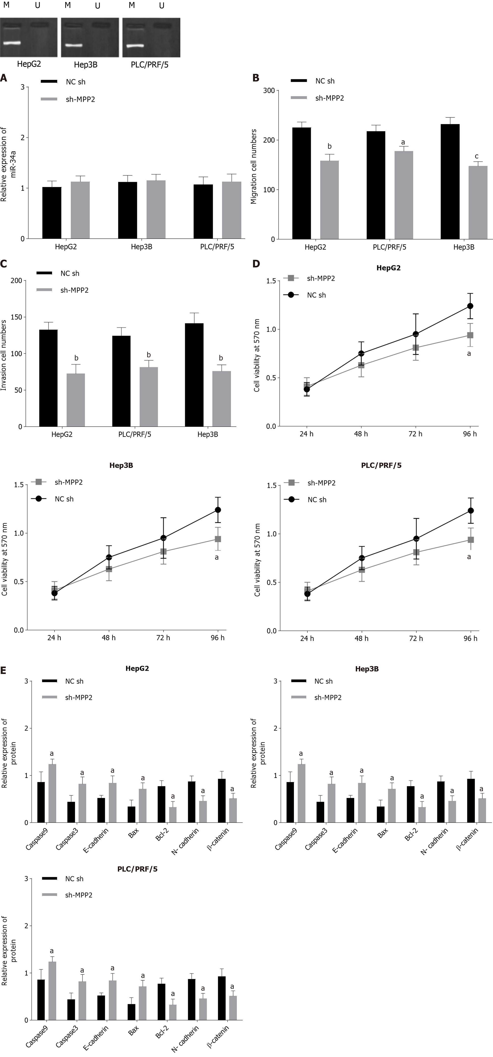
Figure 4 Membrane palmitoylated proteins promote cell apoptosis and inhibits cell proliferation, invasion, and migration.
A: Overexpression of membrane palmitoylated proteins (MPP2) did not affect methylation and the expression level of microRNA 34a (miR-34a); B: MPP2 inhibited cell migration; compared with the negative control-short hairpin (NC-sh) group, a indicates aP < 0.05, bP < 0.01, cP < 0.001; C: MPP2 inhibited cell invasion, compared with the NC-sh group, b indicates bP < 0.01; D: MPP2 inhibited cell activity compared with the NC-sh group, a indicates aP < 0.05; and E: MPP2 inhibited the downregulation of B-cell lymphoma 2 (Bcl-2), N-cadherin and β-catenin expression, but promoted caspase 9, caspase 3, E-cadherin, Bcl-2-associated X protein compared with the NC-sh group, a indicates aP < 0.05).
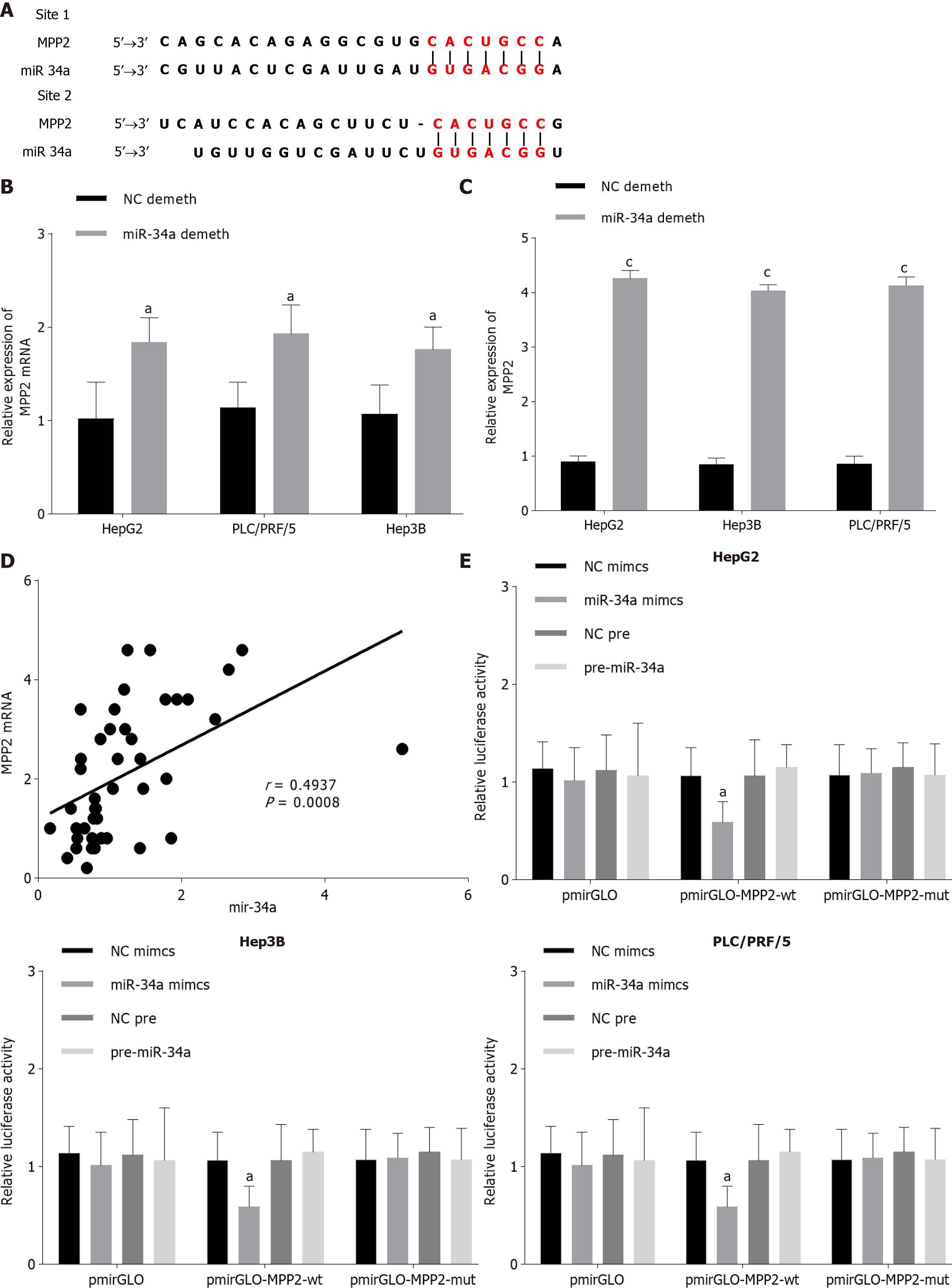
Figure 5 Membrane palmitoylated protein is the target of miR-34a.
A: MicroRNA 34a (miR-34a) and membrane palmitoylated protein (MPP2) mRNA had binding sites; B and C: Effect of miR-34a expression change on MPP2 compared with the negative control (NC) demethylation group, a indicates aP < 0.05; D: miR-34a had a positive correlation with MPP2 mRNA, aP < 0.05, cP < 0.001; and E: Double luciferase reporter gene verified the targeting relationship of miR-34a/MPP2; compared with NC mimics, a indicates aP < 0.05.
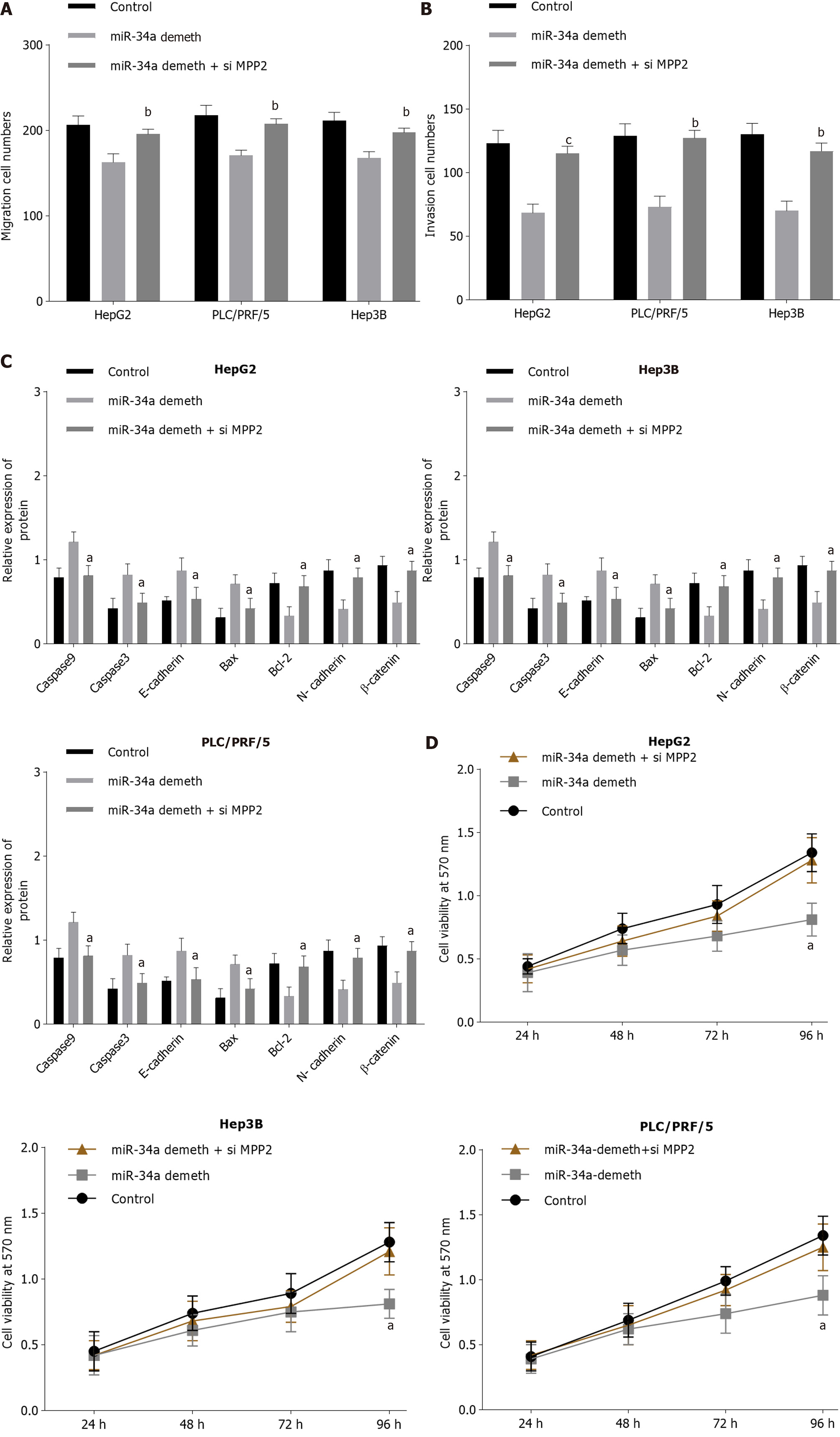
Figure 6 Rescue experiment.
A: Small interfering membrane palmitoylated proteins (si-MPP2) can counteract the effect of microRNA 34a (miR-34a) demethylation on cell migration; B: si-MPP2 counteracts the effect of miR-34a demethylation on cell invasion, bP < 0.01, cP < 0.001; C: si-MPP2 counteracts the effect of miR-34a demethylation on cell activity; and D: si-MPP2 counteracts the effect of miR-34a demethylation on related proteins, a indicates aP < 0.05).
- Citation: Li FY, Fan TY, Zhang H, Sun YM. Demethylation of miR-34a upregulates expression of membrane palmitoylated proteins and promotes the apoptosis of liver cancer cells. World J Gastroenterol 2021; 27(6): 470-486
- URL: https://www.wjgnet.com/1007-9327/full/v27/i6/470.htm
- DOI: https://dx.doi.org/10.3748/wjg.v27.i6.470


