Copyright
©The Author(s) 2021.
World J Gastroenterol. Jun 7, 2021; 27(21): 2664-2680
Published online Jun 7, 2021. doi: 10.3748/wjg.v27.i21.2664
Published online Jun 7, 2021. doi: 10.3748/wjg.v27.i21.2664
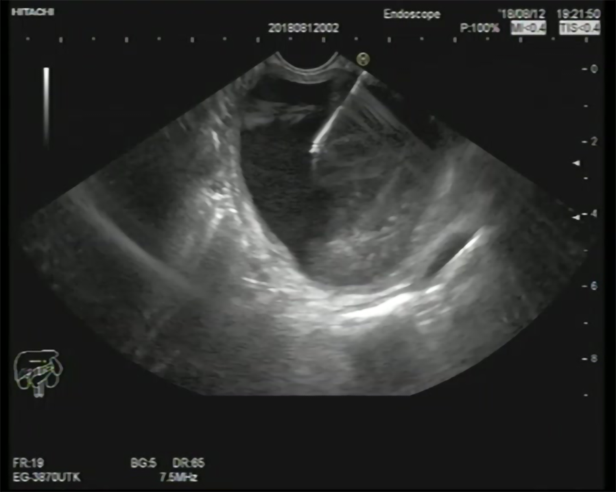
Figure 1
Endoscopic ultrasonography-guided fine-needle aspiration of a pancreatic mucinous cystic neoplasm.
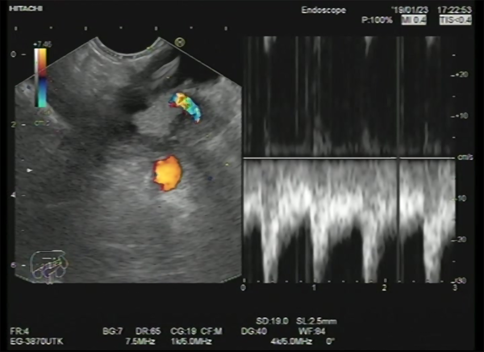
Figure 2
Mucinous cystic neoplasm of pancreatic tail with a mural nodule showing a vessel inside.
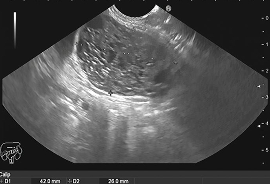
Figure 3
Microcystic serous cystadenoma of the body of the pancreas.
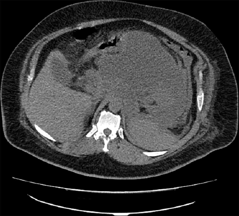
Figure 4
Computed tomography shows inflammatory pancreatic pseudocyst.
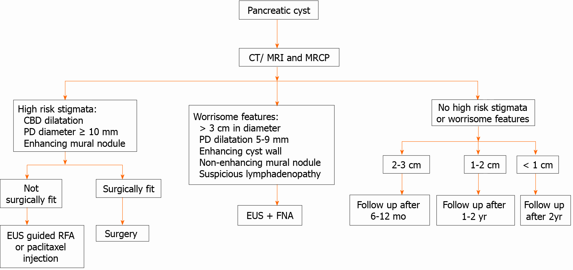
Figure 5 Approach to a patient with a pancreatic cyst.
CT: Computed tomography; MRI: Magnetic resonance imaging; MRCP: Magnetic resonance cholangiopancreatography; CBD: Common bile duct; PD: Pancreatic duct; EUS: Endoscopic ultrasonography; RFA: Radiofrequency ablation; FNA: Fine-needle aspiration.
- Citation: Okasha HH, Awad A, El-meligui A, Ezzat R, Aboubakr A, AbouElenin S, El-Husseiny R, Alzamzamy A. Cystic pancreatic lesions, the endless dilemma. World J Gastroenterol 2021; 27(21): 2664-2680
- URL: https://www.wjgnet.com/1007-9327/full/v27/i21/2664.htm
- DOI: https://dx.doi.org/10.3748/wjg.v27.i21.2664


