Copyright
©The Author(s) 2021.
World J Gastroenterol. Apr 7, 2021; 27(13): 1354-1361
Published online Apr 7, 2021. doi: 10.3748/wjg.v27.i13.1354
Published online Apr 7, 2021. doi: 10.3748/wjg.v27.i13.1354
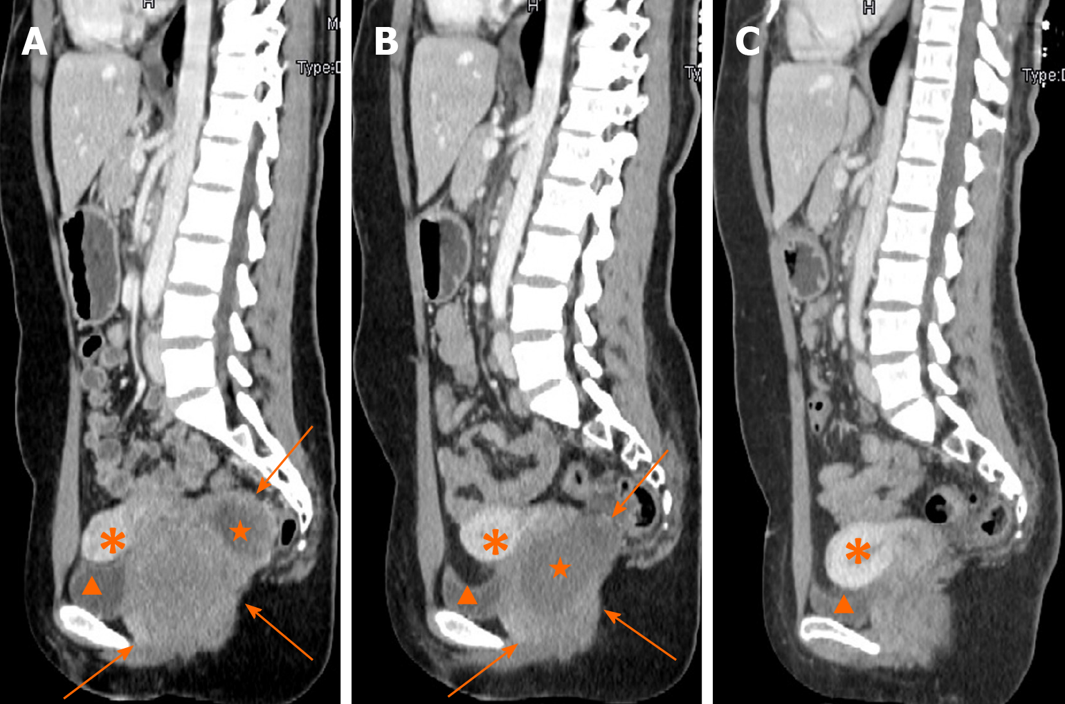
Figure 1 Contrast-enhanced computed tomography in sagittal section.
A: A rectovaginal space mass of 8.0 cm in maximum diameter with non-enhancing liquefaction necrosis at the time of admission; B: The mass significantly shrunk to 6.4 cm in maximum diameter after 5 mo of imatinib treatment and non-enhanced liquefaction area enlarged clearly; C: No obvious sign of tumor recurrence in rectovaginal space 15 mo after tumor resection. Arrows: The mass; pentagrams: Liquefaction necrosis area; arrow heads: Bladder; asterisks: Uterus.
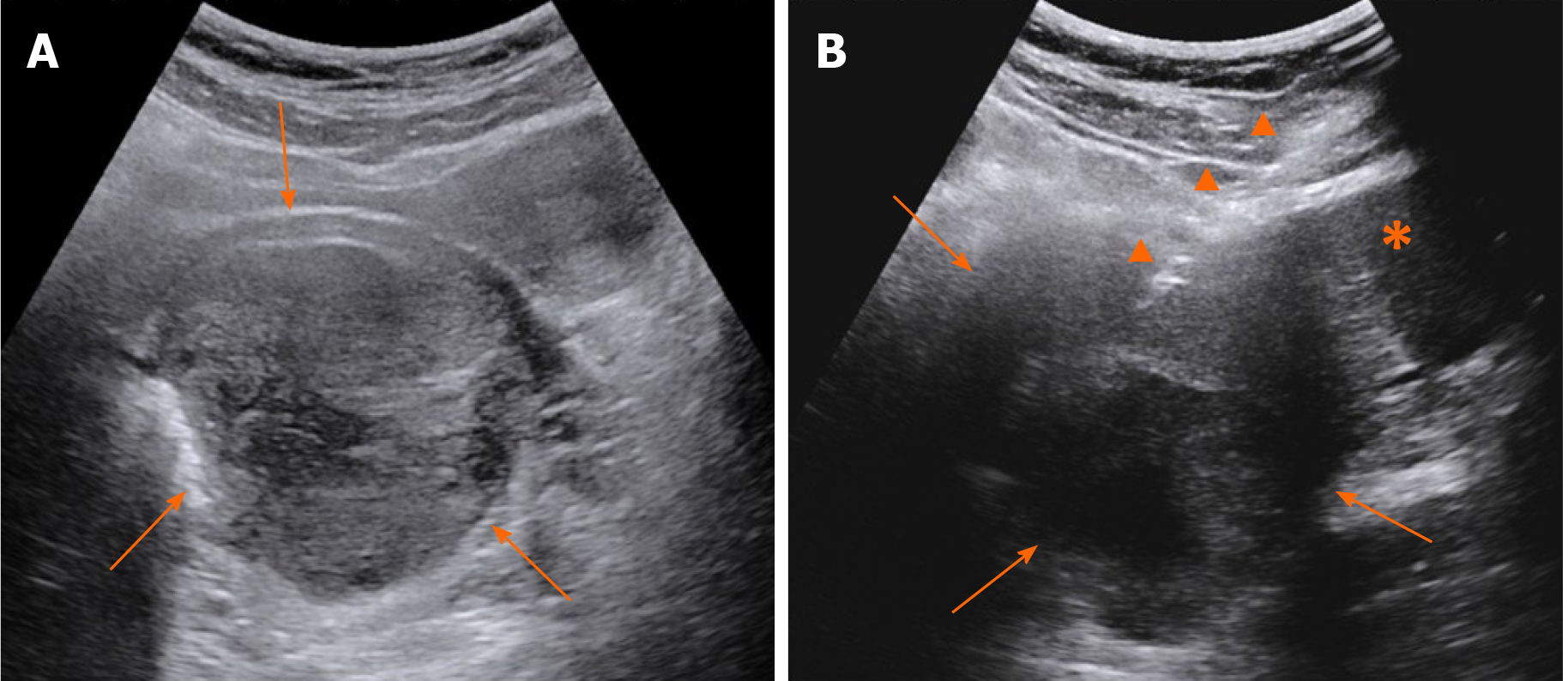
Figure 2 Grey scale ultrasound.
A: A mass was demonstrated inside the rectovaginal space in the grey scale images; B: Transabdominal core needle biopsy of the mass. Arrows: The mass; arrow heads: Core needle; asterisk: Uterus cervix.
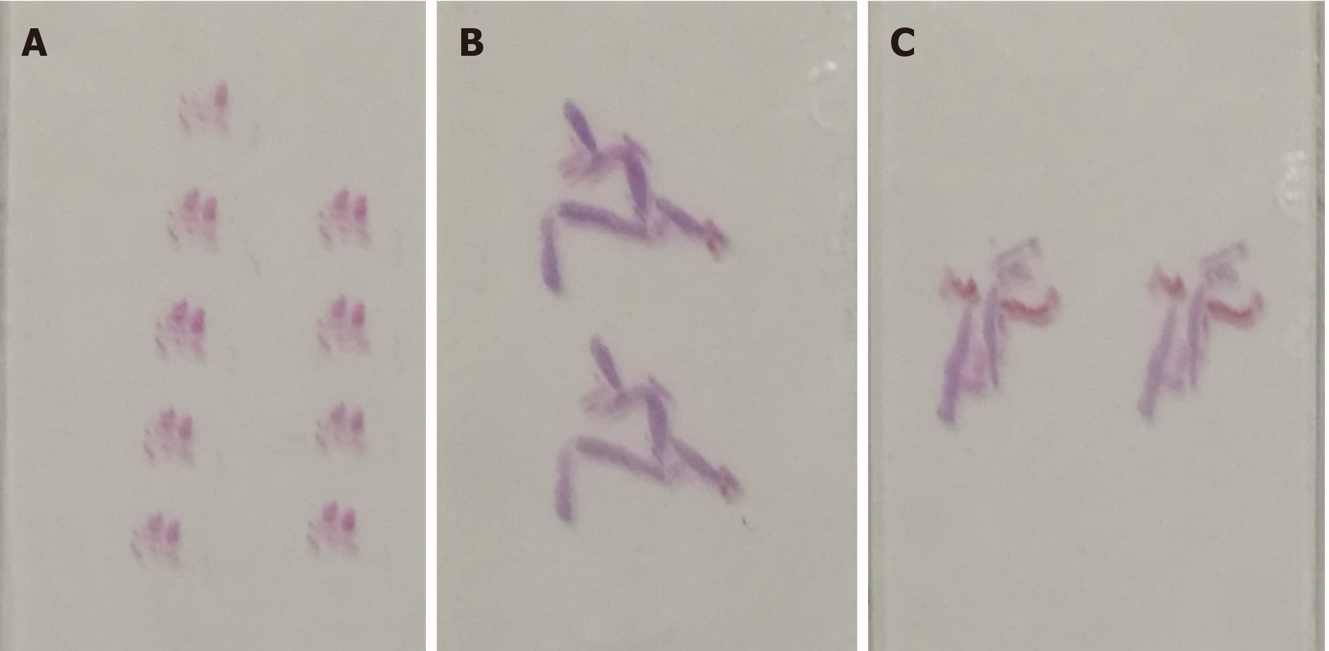
Figure 3 Slide samples.
A: The shattered slide samples from transabdominal ultrasound-guided biopsy; B and C: The slide samples from transperineal biopsy guided by endorectal ultrasound, which were abundant and intact.
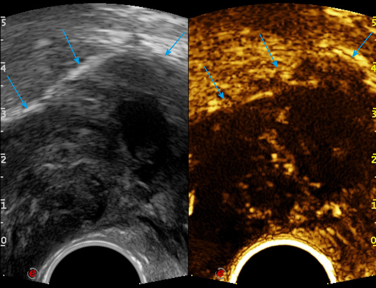
Figure 4 Endorectal ultrasound performed with the convex probe of TRT33.
Liquefaction necrosis and cellular appearance were found inside the tumor in transverse sections, and the solid part of the tumor was either hypo- or iso-enhancing. Arrow: Rectal stromal tumor.
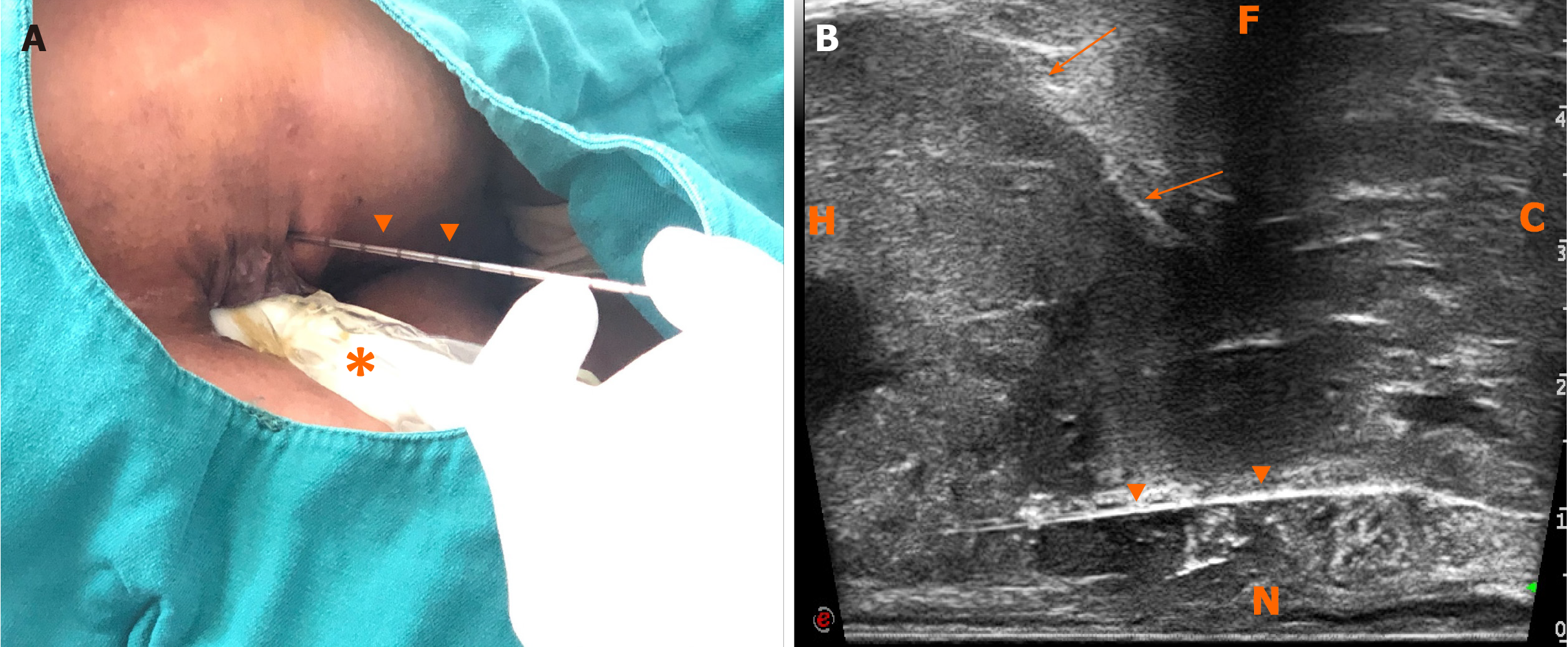
Figure 5 Freehand transperineal biopsy of the rectal subepithelial mass guided by endorectal ultrasound.
A: The patient in left lateral decubitus, and the freehand transperineal biopsy was performed, guided by endorectal ultrasound (ERUS); B: Longitudinal sectional image of transperineal core needle biopsy guided by ERUS obtained with the linear probe of TRT33. H: Head; C: Caudal; N: Near field; F: Far field; arrows: Rectal stromal tumor; triangular arrowheads: Core needle; asterisk: TRT33 probe.
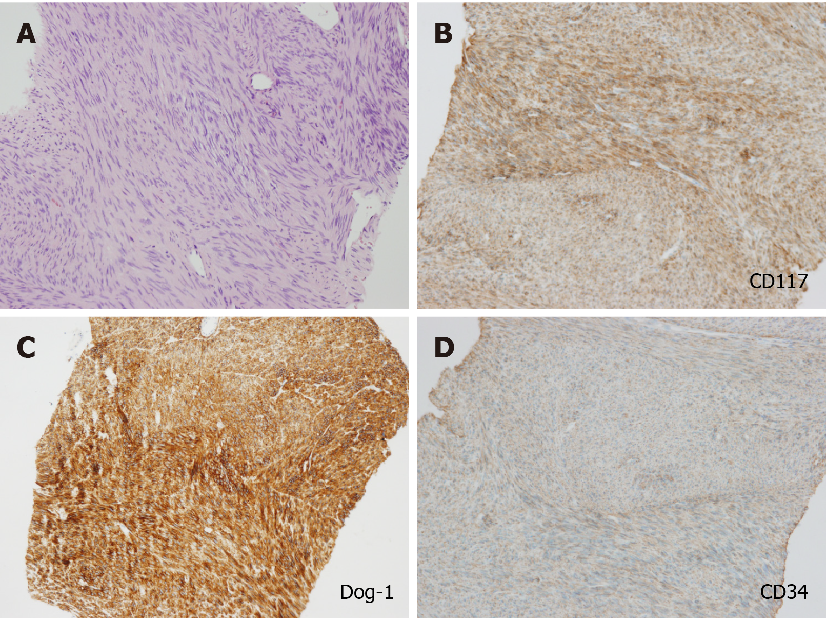
Figure 6 Pathological results.
A: A mass consisting of spindle-shaped cells in hematoxylin and eosin staining; B-D: Immunohistochemical staining of the rectal stromal tumor: CD117, Dog-1, and CD34 were strongly positive (low power field).
- Citation: Zhang Q, Zhao JY, Zhuang H, Lu CY, Yao J, Luo Y, Yu YY. Transperineal core-needle biopsy of a rectal subepithelial lesion guided by endorectal ultrasound after contrast-enhanced ultrasound: A case report. World J Gastroenterol 2021; 27(13): 1354-1361
- URL: https://www.wjgnet.com/1007-9327/full/v27/i13/1354.htm
- DOI: https://dx.doi.org/10.3748/wjg.v27.i13.1354


