Copyright
©The Author(s) 2021.
World J Gastroenterol. Apr 7, 2021; 27(13): 1354-1361
Published online Apr 7, 2021. doi: 10.3748/wjg.v27.i13.1354
Published online Apr 7, 2021. doi: 10.3748/wjg.v27.i13.1354
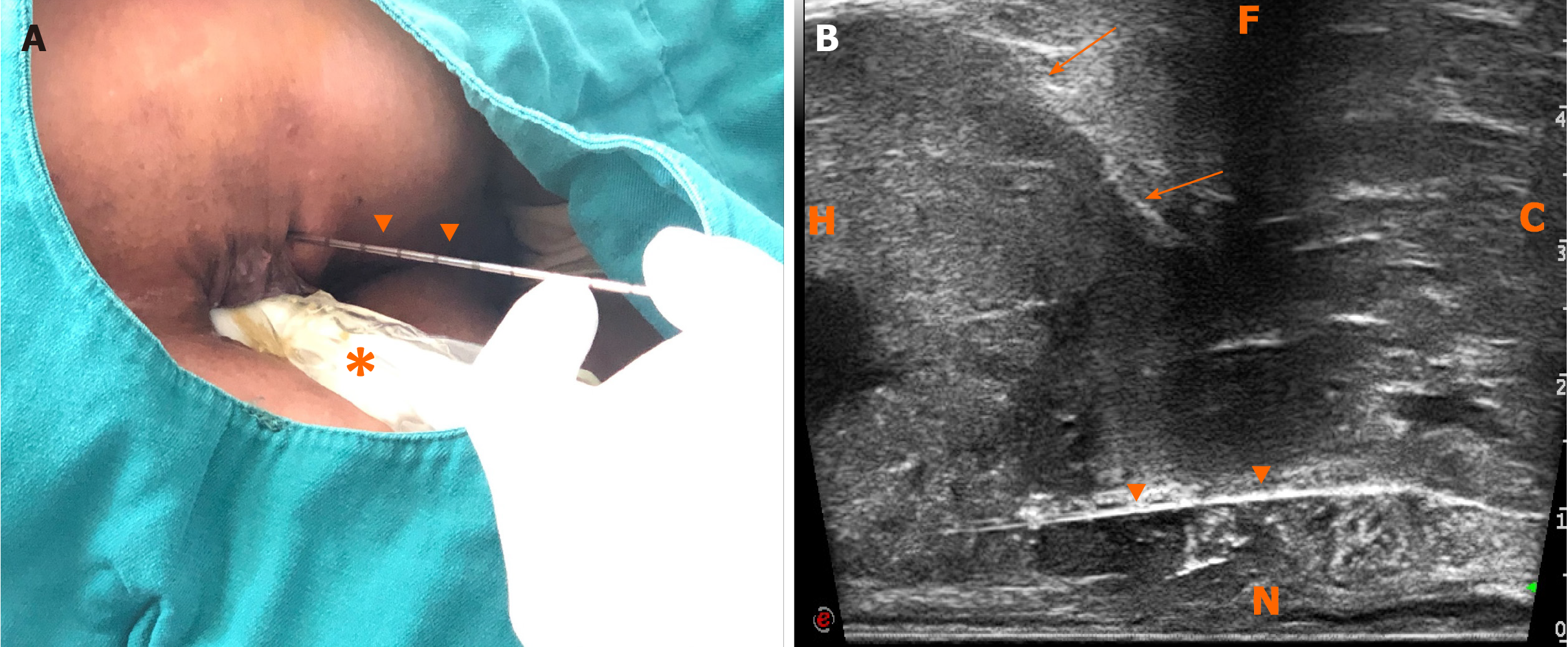
Figure 5 Freehand transperineal biopsy of the rectal subepithelial mass guided by endorectal ultrasound.
A: The patient in left lateral decubitus, and the freehand transperineal biopsy was performed, guided by endorectal ultrasound (ERUS); B: Longitudinal sectional image of transperineal core needle biopsy guided by ERUS obtained with the linear probe of TRT33. H: Head; C: Caudal; N: Near field; F: Far field; arrows: Rectal stromal tumor; triangular arrowheads: Core needle; asterisk: TRT33 probe.
- Citation: Zhang Q, Zhao JY, Zhuang H, Lu CY, Yao J, Luo Y, Yu YY. Transperineal core-needle biopsy of a rectal subepithelial lesion guided by endorectal ultrasound after contrast-enhanced ultrasound: A case report. World J Gastroenterol 2021; 27(13): 1354-1361
- URL: https://www.wjgnet.com/1007-9327/full/v27/i13/1354.htm
- DOI: https://dx.doi.org/10.3748/wjg.v27.i13.1354


