Copyright
©The Author(s) 2021.
World J Gastroenterol. Apr 7, 2021; 27(13): 1354-1361
Published online Apr 7, 2021. doi: 10.3748/wjg.v27.i13.1354
Published online Apr 7, 2021. doi: 10.3748/wjg.v27.i13.1354
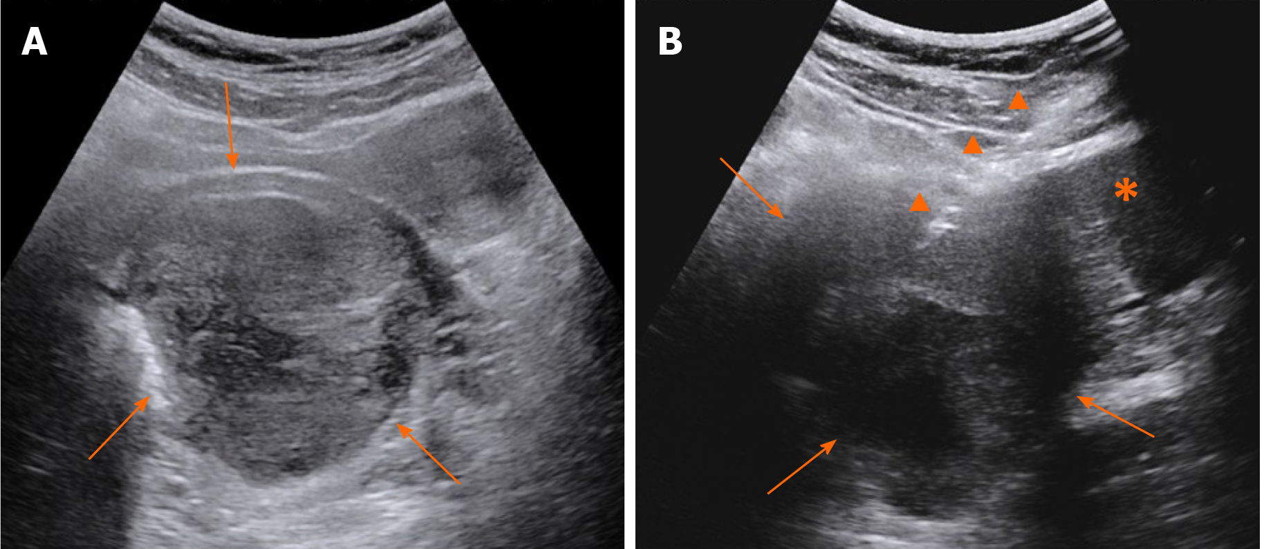
Figure 2 Grey scale ultrasound.
A: A mass was demonstrated inside the rectovaginal space in the grey scale images; B: Transabdominal core needle biopsy of the mass. Arrows: The mass; arrow heads: Core needle; asterisk: Uterus cervix.
- Citation: Zhang Q, Zhao JY, Zhuang H, Lu CY, Yao J, Luo Y, Yu YY. Transperineal core-needle biopsy of a rectal subepithelial lesion guided by endorectal ultrasound after contrast-enhanced ultrasound: A case report. World J Gastroenterol 2021; 27(13): 1354-1361
- URL: https://www.wjgnet.com/1007-9327/full/v27/i13/1354.htm
- DOI: https://dx.doi.org/10.3748/wjg.v27.i13.1354


