Copyright
©The Author(s) 2020.
World J Gastroenterol. Dec 14, 2020; 26(46): 7416-7424
Published online Dec 14, 2020. doi: 10.3748/wjg.v26.i46.7416
Published online Dec 14, 2020. doi: 10.3748/wjg.v26.i46.7416
Figure 1 Region of interest in the liver in volunteers and in patients with focal liver lesions using virtual touch tissue quantification.
A: Region of interest (with fixed size 10 mm × 5 mm) for the liver in volunteers was placed at a depth of 4-6 cm, and tubular structures, such as portal veins, hepatic veins and intrahepatic bile ducts were carefully avoided; B: Region of interest for patients was placed inside the targeted focal liver lesion (a hemangioma shown here).
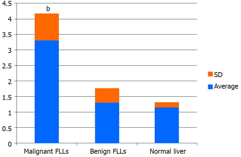
Figure 2 Comparison of maximum elasticity among normal livers, benign focal liver lesions, and malignant focal liver lesions.
bP < 0.01 compared with maximum elasticity (Emax) of benign focal liver lesions (FLLs) or normal livers. Emax of malignant FLLs were statistically significantly higher compared with Emax of benign FLLs and normal livers. There was no statistically significant difference between Emax values of the benign FLLs and normal livers.
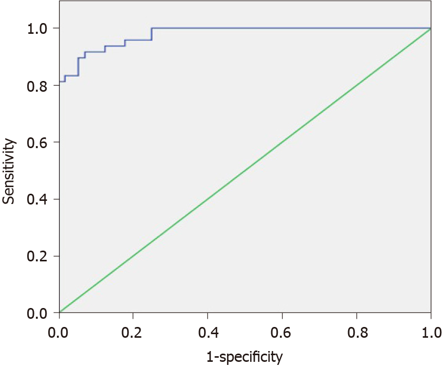
Figure 3 Receiver operating characteristic curves of maximum elasticity for malignant and benign focal liver lesions.
- Citation: Zhang HP, Gu JY, Bai M, Li F, Zhou YQ, Du LF. Value of shear wave elastography with maximal elasticity in differentiating benign and malignant solid focal liver lesions. World J Gastroenterol 2020; 26(46): 7416-7424
- URL: https://www.wjgnet.com/1007-9327/full/v26/i46/7416.htm
- DOI: https://dx.doi.org/10.3748/wjg.v26.i46.7416


