Copyright
©The Author(s) 2018.
World J Gastroenterol. Jul 28, 2018; 24(28): 3192-3197
Published online Jul 28, 2018. doi: 10.3748/wjg.v24.i28.3192
Published online Jul 28, 2018. doi: 10.3748/wjg.v24.i28.3192
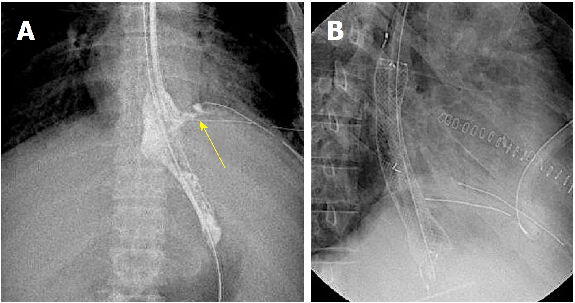
Figure 1 Fluoroscopic image.
Leakage of the contrast agent from the suture site was confirmed (A, arrow); and a fully covered self-expandable metallic stent was placed in the leakage site (B).
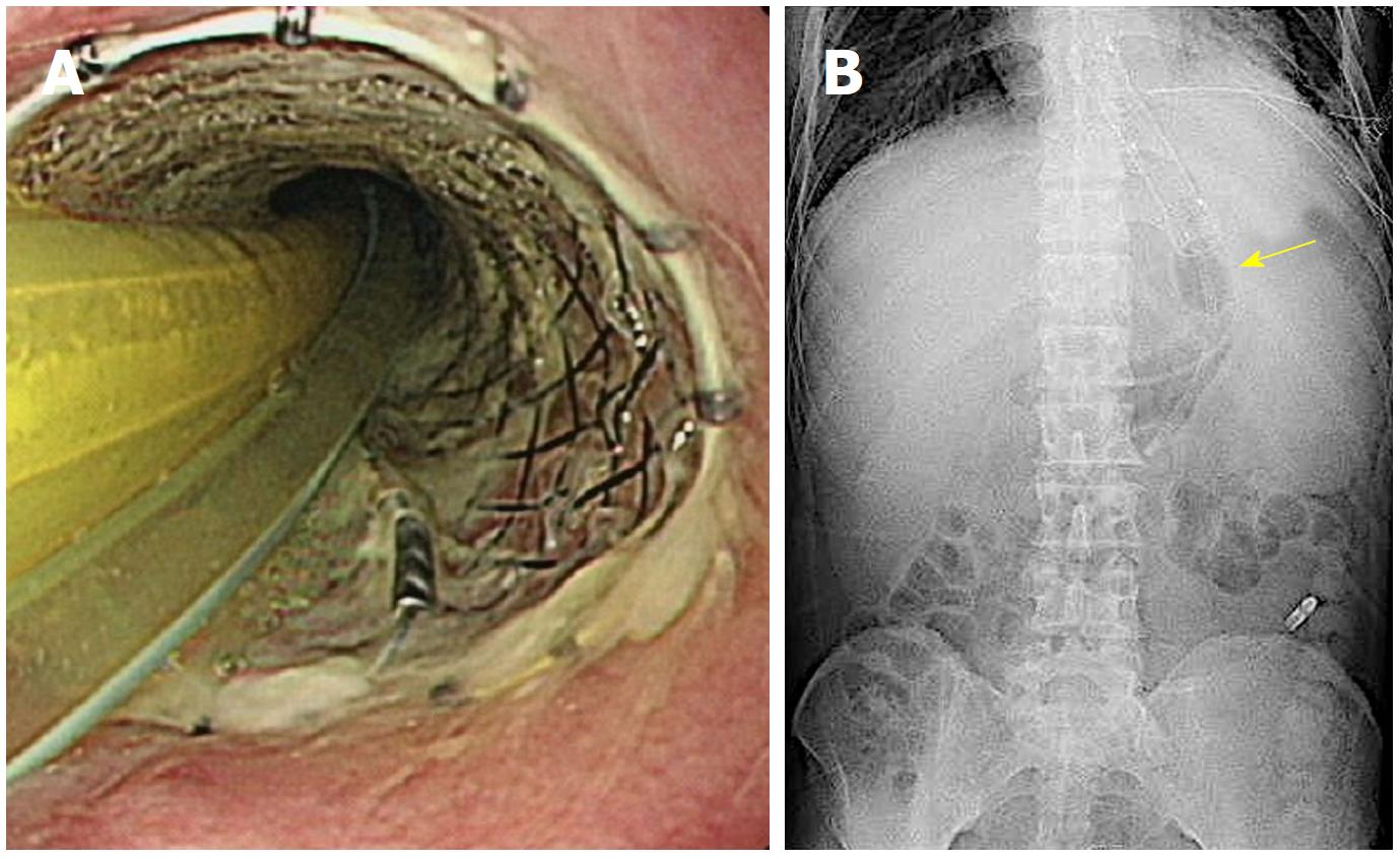
Figure 2 Sengstaken-Blakemore tube and nasoesophageal feeding tube placement.
These tubes were placed by passing through the stent lumen (A); and the Sengstaken-Blakemore tube gastric balloon was inflated slightly larger than the stent diameter (B, arrow) to support the stent.
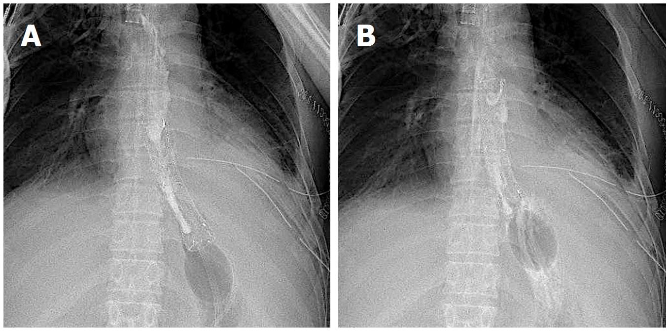
Figure 3 Fluoroscopic image.
Stent migration was confirmed. While supporting the stent with the gastric balloon, the SBT was pulled toward the oral side to correct the position. A: Before repositioning; B: After repositioning. SBT: Sengstaken-Blakemore tube.
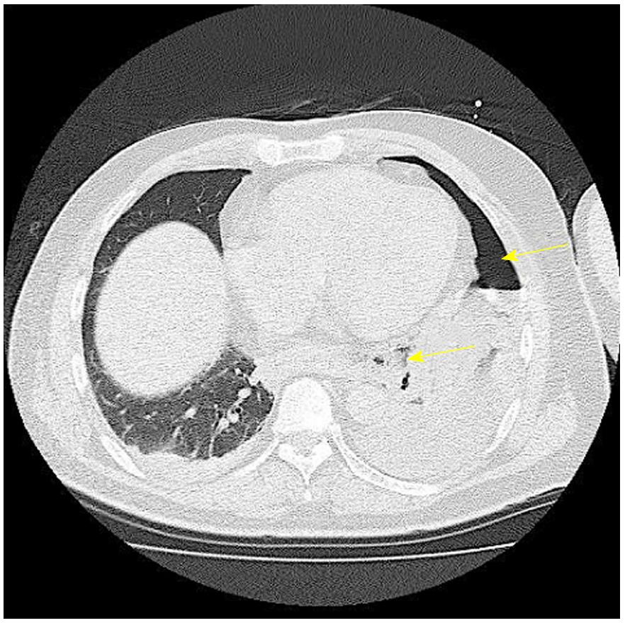
Figure 4 Chest computed tomography scan.
Scan confirmed left pneumothorax (arrow) and mediastinal emphysema (arrow).
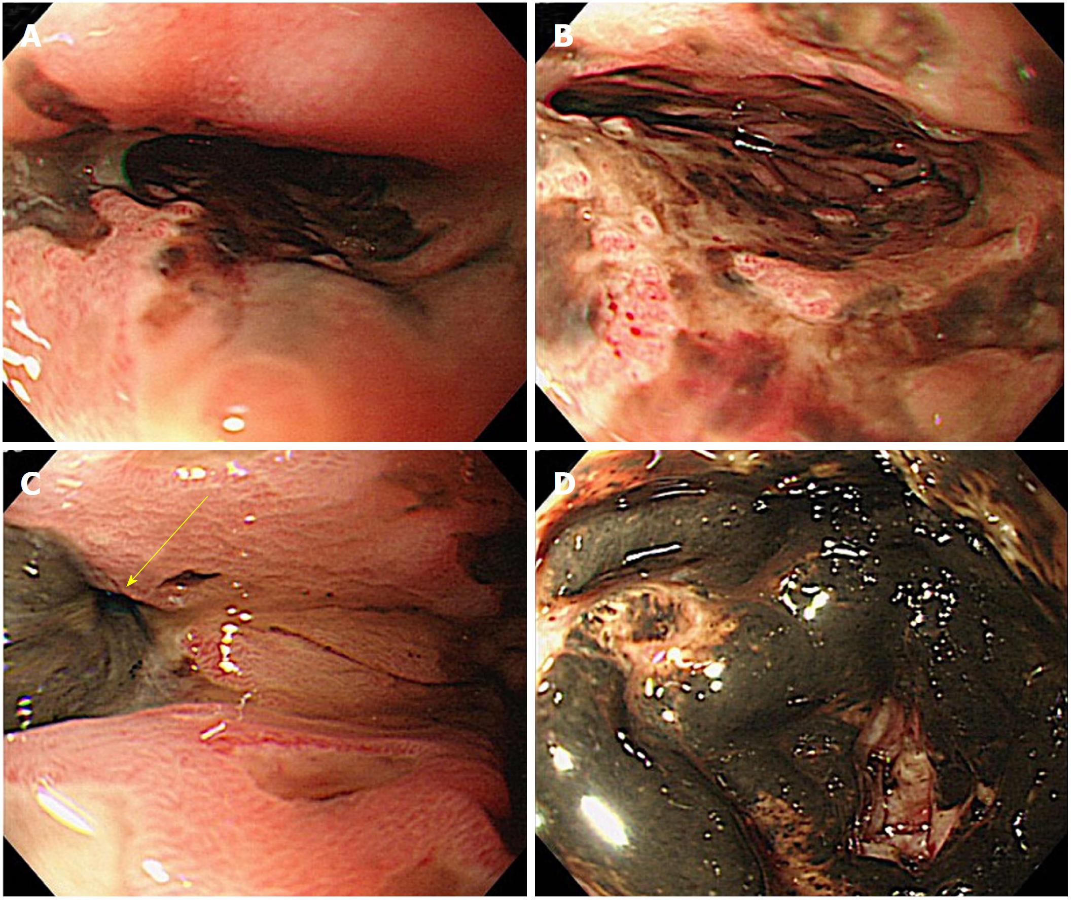
Figure 5 Initial Esophagogastroduodenoscopy findings.
A and B: Black mucosal lesion was confirmed in the middle to lower esophagus; C: Perforation (arrow) due to necrosis was confirmed in the left wall of the lower esophagus; D: There was a black mucosal lesion in the duodenum.
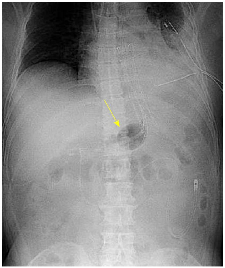
Figure 6 Abdominal radiograph.
The SBT gastric balloon was inflated slightly larger than the stent diameter (arrow) to support the stent. The nasoesophageal feeding tube was positioned in the jejunum. SBT: Sengstaken-Blakemore tube.
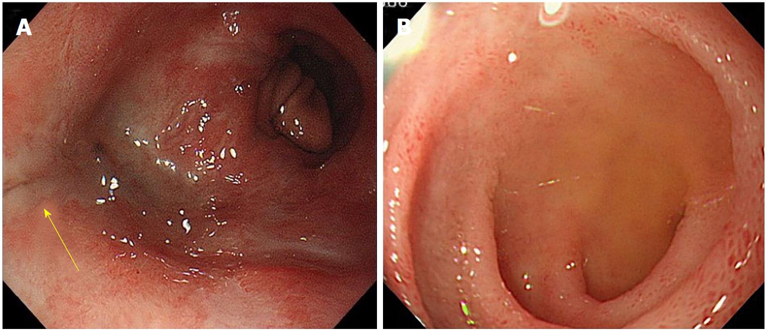
Figure 7 Esophagogastroduodenoscopy findings upon stent removal.
A: Closure of the perforated site (arrow) was confirmed; B: The mucous membrane of the duodenum improved.
- Citation: Sato H, Ishida K, Sasaki S, Kojika M, Endo S, Inoue Y, Sasaki A. Regulating migration of esophageal stents - management using a Sengstaken-Blakemore tube: A case report and review of literature. World J Gastroenterol 2018; 24(28): 3192-3197
- URL: https://www.wjgnet.com/1007-9327/full/v24/i28/3192.htm
- DOI: https://dx.doi.org/10.3748/wjg.v24.i28.3192


