Copyright
©The Author(s) 2018.
World J Gastroenterol. Jul 28, 2018; 24(28): 3192-3197
Published online Jul 28, 2018. doi: 10.3748/wjg.v24.i28.3192
Published online Jul 28, 2018. doi: 10.3748/wjg.v24.i28.3192
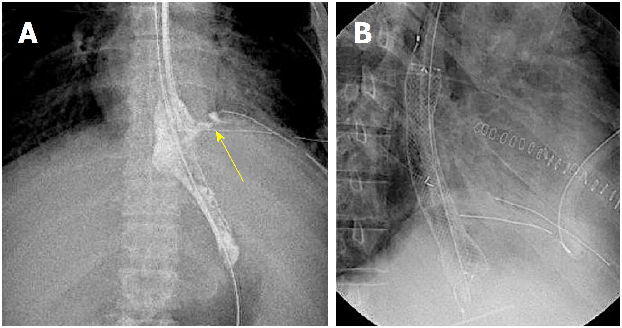
Figure 1 Fluoroscopic image.
Leakage of the contrast agent from the suture site was confirmed (A, arrow); and a fully covered self-expandable metallic stent was placed in the leakage site (B).
- Citation: Sato H, Ishida K, Sasaki S, Kojika M, Endo S, Inoue Y, Sasaki A. Regulating migration of esophageal stents - management using a Sengstaken-Blakemore tube: A case report and review of literature. World J Gastroenterol 2018; 24(28): 3192-3197
- URL: https://www.wjgnet.com/1007-9327/full/v24/i28/3192.htm
- DOI: https://dx.doi.org/10.3748/wjg.v24.i28.3192


