Copyright
©The Author(s) 2018.
World J Gastroenterol. Jul 14, 2018; 24(26): 2915-2920
Published online Jul 14, 2018. doi: 10.3748/wjg.v24.i26.2915
Published online Jul 14, 2018. doi: 10.3748/wjg.v24.i26.2915
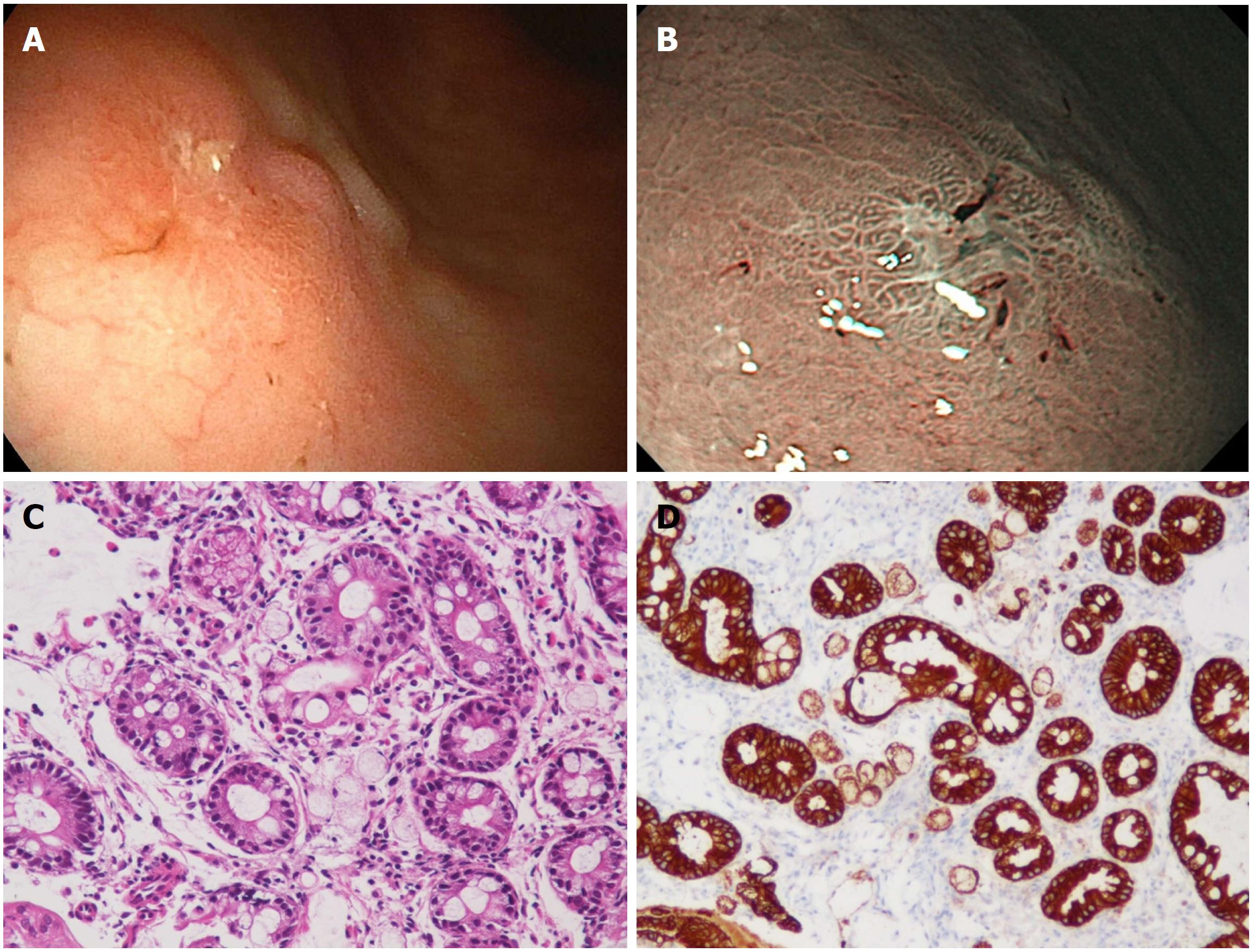
Figure 1 Image from esophagogastroduodenoscopy.
A: Depressed lesion was found at gastric angle of the greater curvature side; B: The narrow band imaging of the EGD showed a relatively demarcated lesion with an irregular microsurface pattern; C: The biopsy specimen from the depressed lesion. Among the glands with intestinal metaplasia, a small number of signet-ring cell carcinoma cells were found (HE; × 200). D: Signet-ring cell carcinoma cells were positive for immunohistochemistry of pan-cytokeratin (× 200). EGD: Esophagogastroduodenoscopy.
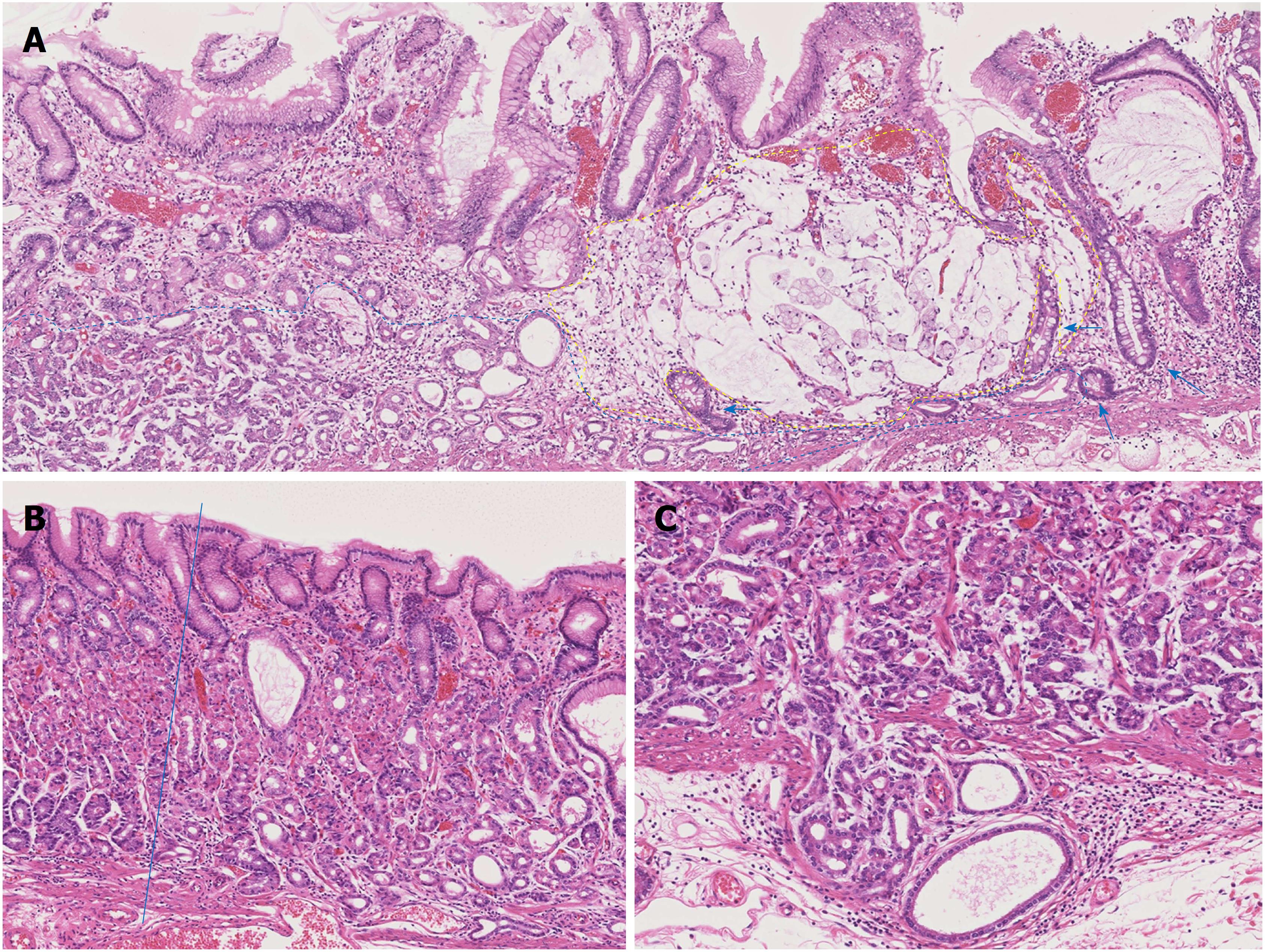
Figure 2 Pathological findings.
A: Representative histological photograph of the specimens of endoscopic submucosal dissection (HE; × 50). Proliferation of gastric adenocarcinoma of the fundic gland type (GA-FG) are observed at the deep layer of the lamina propria mucosae in the left half of the photo (blue dot line: the border of GA-FG). Adjacent to the GA-FG, proliferation of the signet-ring cell carcinoma producing intra- and extracellular mucin is observed in the right half of the photo (yellow dotted line: border of the signet-ring cell carcinoma). Intestinal metaplasia was observed at the mucosa surrounding the signet-ring cell carcinoma (arrows); B: Structure and differentiation toward the surfaces of the fundic gland were significantly disturbed at the GA-FG compared to the normal fundic glands (HE; × 50). The blue line is the border of the GA-FG and the normal fundic glands. The mucosal surface was covered with non-neoplastic foveolar epithelium. Intestinal metaplasia cannot be observed in this photo; C: GA-FG invaded into the submucosal layer (HE; × 100).
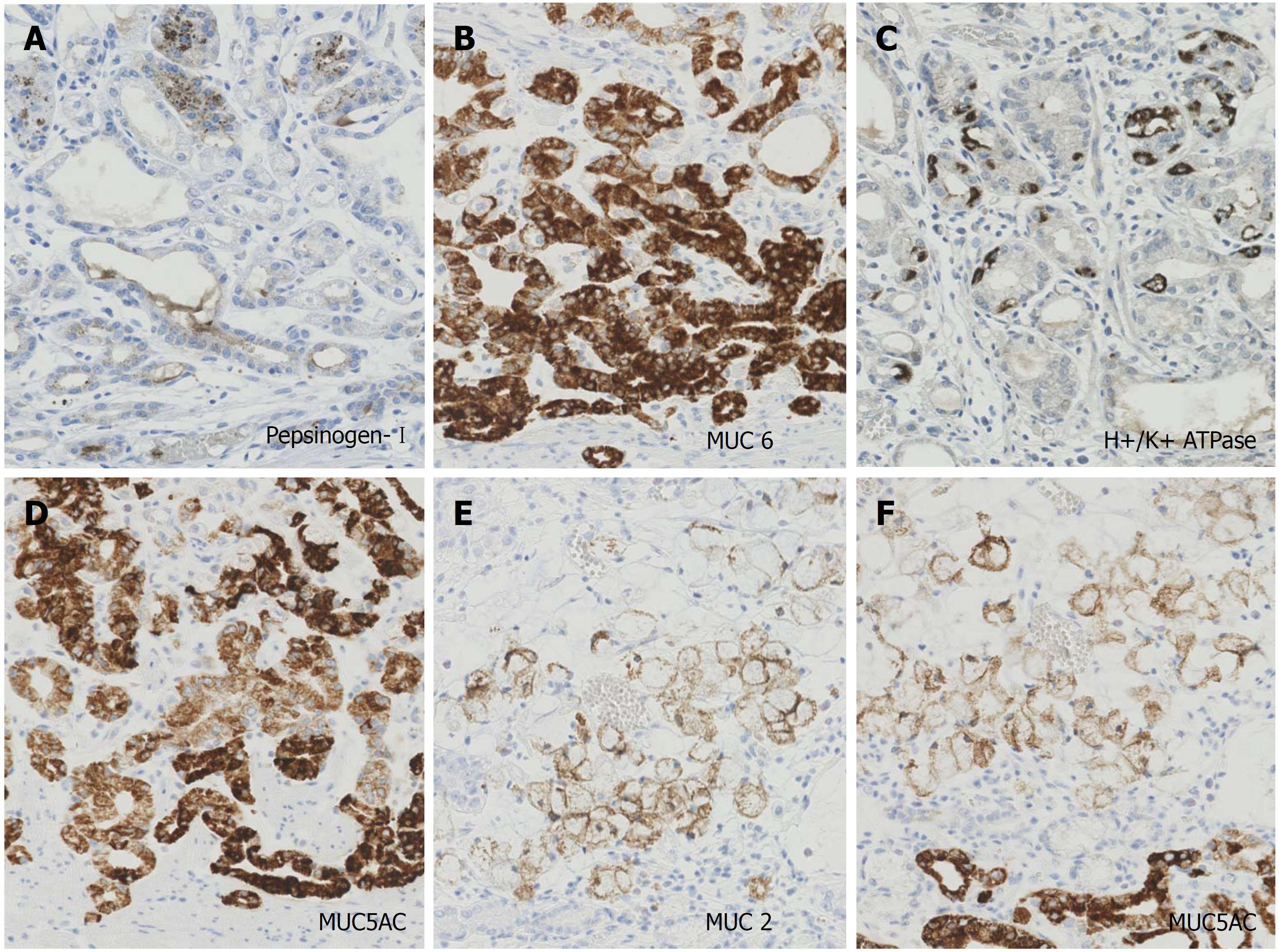
Figure 3 Photographs of immunohistochemistry.
The magnifications of all photographs are × 200. The tumor cells of GA-FG expressed focally (30%) pepsinogen-I (A), diffusely MUC6 (B), scattered (5%) H+/K+ ATPase (C), and diffusely MUC5AC (D). The tumor cells of the signet-ring cell carcinoma diffusely expressed MUC 2 (E) and MUC 5AC (F). GA-FG: Gastric adenocarcinoma of fundic gland type.
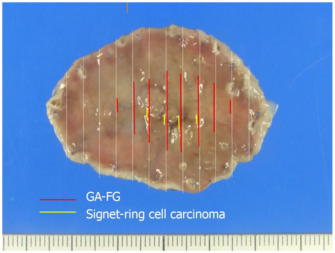
Figure 4 Mapping of the endoscopic submucosal dissection specimen based on histology.
GA-FG distributed at a slightly depressed lesion measuring 28 mm × 14 mm (red line) and signet-ring cell carcinoma distributed at a deeper depressed lesion measuring 12 mm × 3 mm in the slightly depressed lesion (yellow line). GA-FG: Gastric adenocarcinoma of fundic gland type.
- Citation: Kai K, Satake M, Tokunaga O. Gastric adenocarcinoma of fundic gland type with signet-ring cell carcinoma component: A case report and review of the literature. World J Gastroenterol 2018; 24(26): 2915-2920
- URL: https://www.wjgnet.com/1007-9327/full/v24/i26/2915.htm
- DOI: https://dx.doi.org/10.3748/wjg.v24.i26.2915


