Copyright
©The Author(s) 2018.
World J Gastroenterol. Jul 14, 2018; 24(26): 2915-2920
Published online Jul 14, 2018. doi: 10.3748/wjg.v24.i26.2915
Published online Jul 14, 2018. doi: 10.3748/wjg.v24.i26.2915
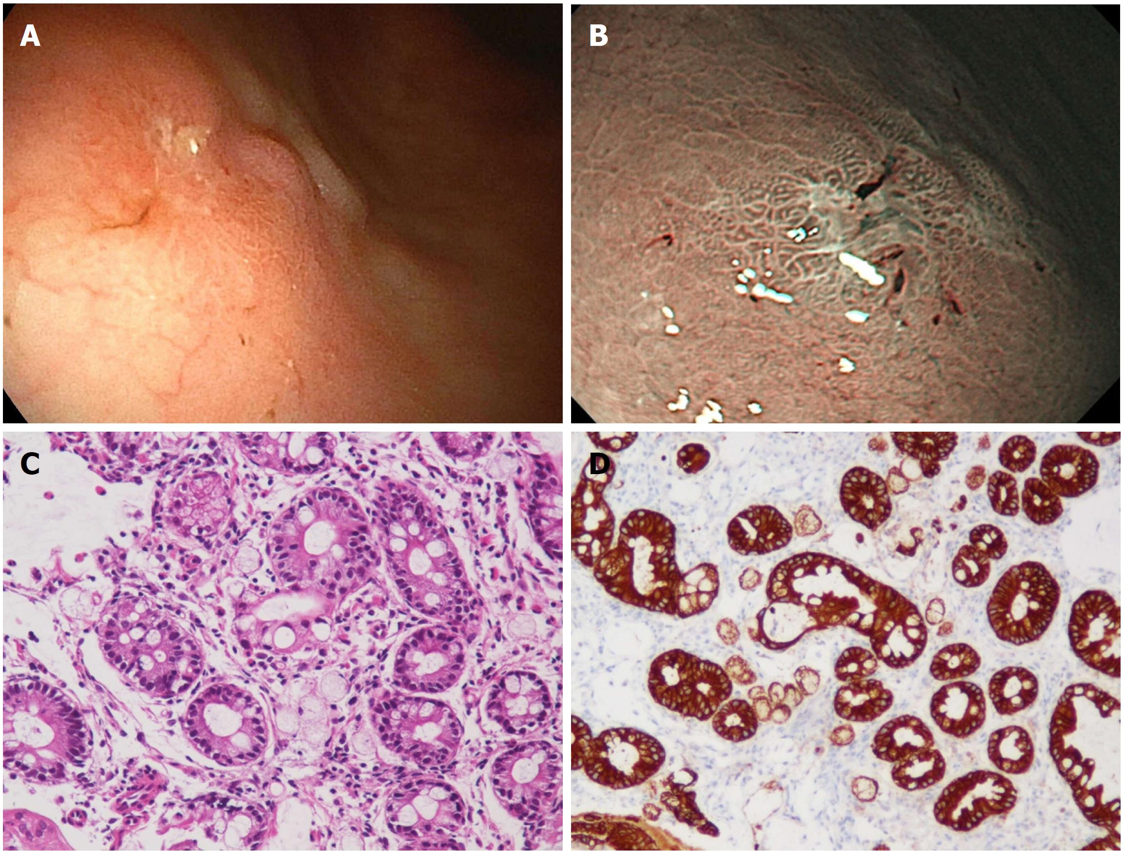
Figure 1 Image from esophagogastroduodenoscopy.
A: Depressed lesion was found at gastric angle of the greater curvature side; B: The narrow band imaging of the EGD showed a relatively demarcated lesion with an irregular microsurface pattern; C: The biopsy specimen from the depressed lesion. Among the glands with intestinal metaplasia, a small number of signet-ring cell carcinoma cells were found (HE; × 200). D: Signet-ring cell carcinoma cells were positive for immunohistochemistry of pan-cytokeratin (× 200). EGD: Esophagogastroduodenoscopy.
- Citation: Kai K, Satake M, Tokunaga O. Gastric adenocarcinoma of fundic gland type with signet-ring cell carcinoma component: A case report and review of the literature. World J Gastroenterol 2018; 24(26): 2915-2920
- URL: https://www.wjgnet.com/1007-9327/full/v24/i26/2915.htm
- DOI: https://dx.doi.org/10.3748/wjg.v24.i26.2915


