Copyright
©The Author(s) 2018.
World J Gastroenterol. Jul 14, 2018; 24(26): 2915-2920
Published online Jul 14, 2018. doi: 10.3748/wjg.v24.i26.2915
Published online Jul 14, 2018. doi: 10.3748/wjg.v24.i26.2915
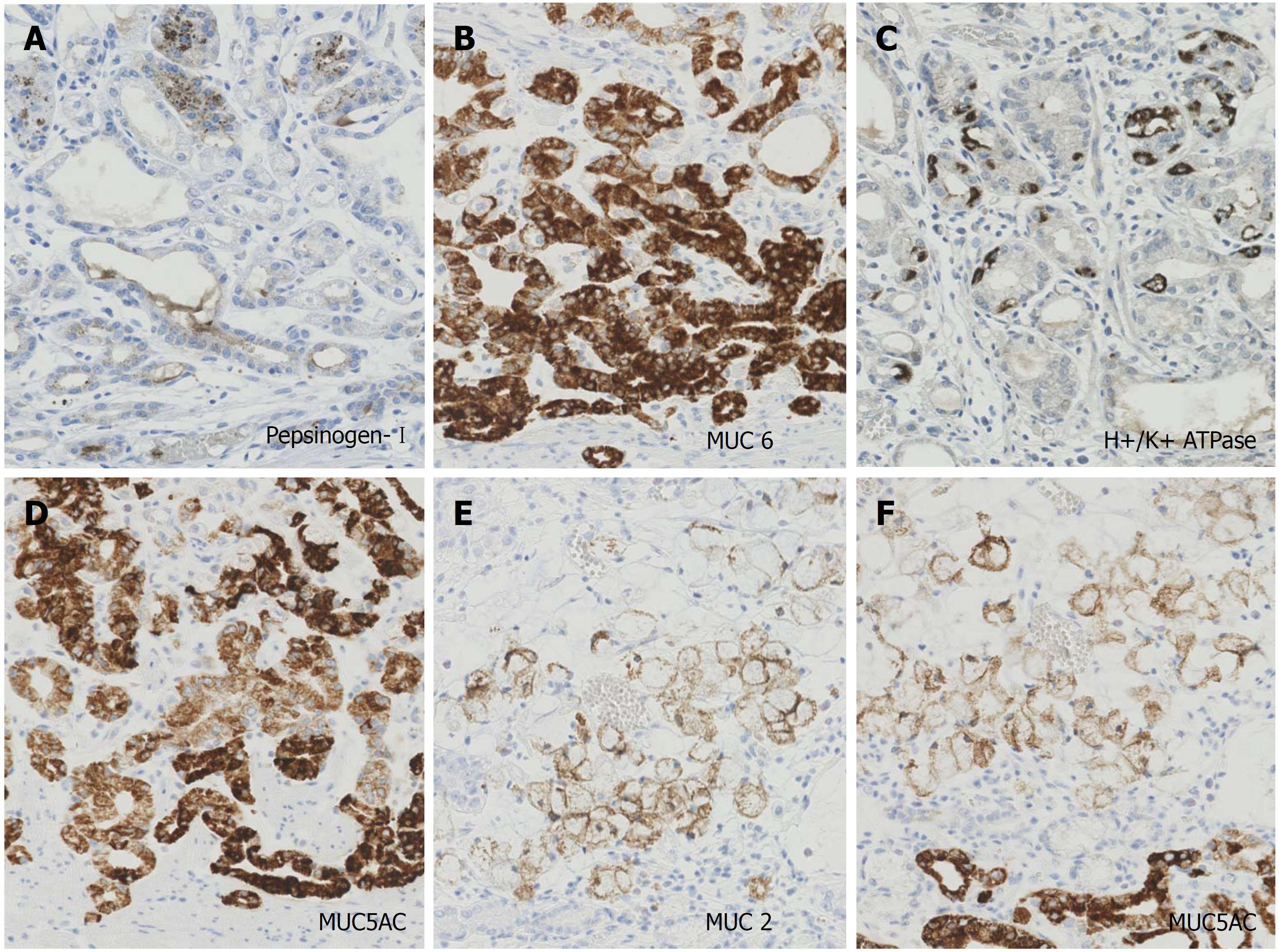
Figure 3 Photographs of immunohistochemistry.
The magnifications of all photographs are × 200. The tumor cells of GA-FG expressed focally (30%) pepsinogen-I (A), diffusely MUC6 (B), scattered (5%) H+/K+ ATPase (C), and diffusely MUC5AC (D). The tumor cells of the signet-ring cell carcinoma diffusely expressed MUC 2 (E) and MUC 5AC (F). GA-FG: Gastric adenocarcinoma of fundic gland type.
- Citation: Kai K, Satake M, Tokunaga O. Gastric adenocarcinoma of fundic gland type with signet-ring cell carcinoma component: A case report and review of the literature. World J Gastroenterol 2018; 24(26): 2915-2920
- URL: https://www.wjgnet.com/1007-9327/full/v24/i26/2915.htm
- DOI: https://dx.doi.org/10.3748/wjg.v24.i26.2915


