Copyright
©The Author(s) 2018.
World J Gastroenterol. Jan 7, 2018; 24(1): 150-156
Published online Jan 7, 2018. doi: 10.3748/wjg.v24.i1.150
Published online Jan 7, 2018. doi: 10.3748/wjg.v24.i1.150
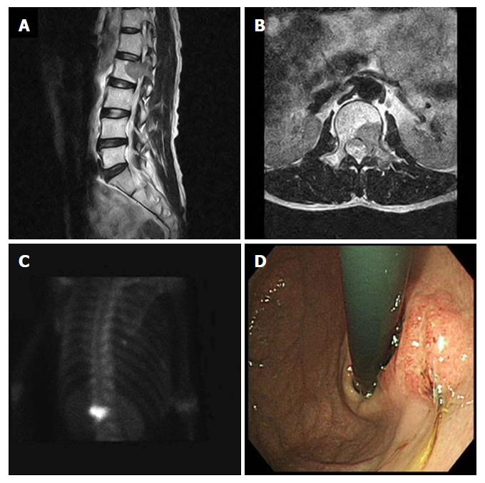
Figure 1 Radiologic findings of the 2nd lumbar vertebra and the endoscopic finding of gastric cancer.
A and B: Bone magnetic resonance imaging showing bone marrow signal change and soft tissue formation at the 2nd lumbar vertebra that extended to the transverse process and the back muscle; C: Bone scan demonstrating 99m Tc-HDP 25mCi uptake in the 2nd lumbar vertebra; D: Endoscopy revealing a 3-cm ulceroinfiltrative mass on the lesser curvature of the high body of the remnant stomach.
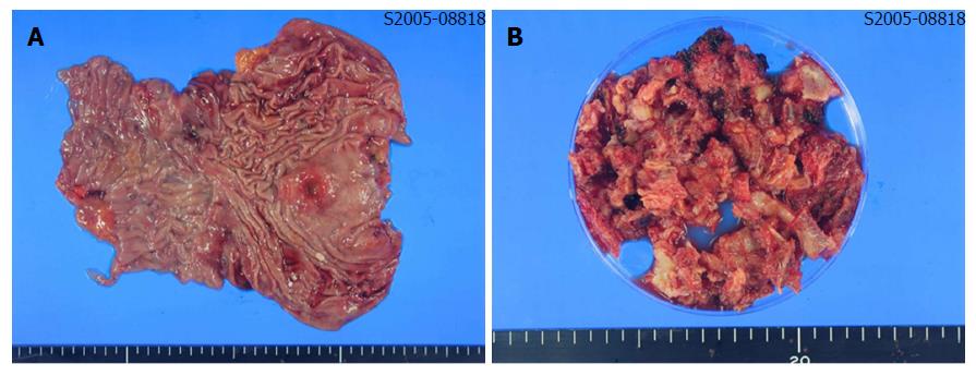
Figure 2 Macroscopic findings of resected stomach and metastatic tumor of bone.
A: A 3-cm ulceroinfiltrating mass was found on the lesser curvature side of stomach. It was 4 cm distant from the proximal resection margin, and 11 cm distant from the distal resection margin; B: About 100 cc of bone and soft tissues were resected from the lumbar spine.
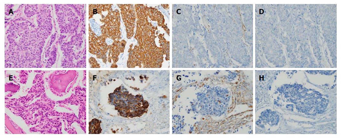
Figure 3 Histopathological and immunohistochemical findings of stomach cancer (A-D) and metastatic bone lesion (E and F).
A: The stomach tumor consisted of solid nests of poorly differentiated tumor cells, with ovoid nuclei and indistinct cytoplasm (hematoxylin-eosin, × 400); B-D: Immunohistochemistry showed that the tumor cells exhibited diffuse immunoreactivity for cytokeratin AE1/AE3 (B: × 400), but were negative for vimentin (C: × 400) and synaptophysin (D: × 400), which supported the diagnosis of poorly differentiated adenocarcinoma; E: The bone tumors were identified as poorly differentiated tumors (hematoxylin-eosin, × 400), with histologic and immunohistochemical features identical to those of the carcinoma of the stomach that were positive for cytokeratin AE1/AE3 (F: × 400) and negative for vimentin (G: × 400) and synaptophysin (H: × 400).
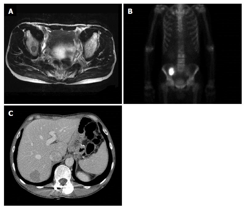
Figure 4 Findings of bone magnetic resonance imaging, bone scan and abdomen computed tomography after 25-mo of follow-up.
A and B: Right ileum bone metastasis; C: Liver metastasis.
- Citation: Choi YJ, Kim DH, Han HS, Han JH, Son SM, Kim DS, Yun HY. Long-term survival after gastrectomy and metastasectomy for gastric cancer with synchronous bone metastasis. World J Gastroenterol 2018; 24(1): 150-156
- URL: https://www.wjgnet.com/1007-9327/full/v24/i1/150.htm
- DOI: https://dx.doi.org/10.3748/wjg.v24.i1.150


